
II. Differential Diagnosis: Acute Vision Loss based on pain
- Painful Vision Loss or Blurred Vision
- Eye Injury
- Corneal Ulcer
- Acute Angle-Closure Glaucoma (esp. if Intraocular Pressure >60 mmHg)
- Iritis and Uveitis (anterior chamber exudates)
- Endophthalmitis (vitreous exudates)
- Painless (or Minimal or variable pain) Vision Loss or Blurred Vision
- Optic Neuritis, retrobulbar Optic Neuritis or Papillitis (associated with Multiple Sclerosis)
- Retinal Detachment
- Ocular tumor
- Central Retinal Artery Occlusion (pale fundus with cherry red Macula)
- Acute Maculopathy
- Pseudotumor Cerebri (or other cause of Increased Intracranial Pressure)
- Associated with Headache
- Transient Monocular Blindness (Amaurosis Fugax)
- Retinal Hemorrhage
III. Differential Diagnosis: Acute Unilateral Vision Loss
- Transient
- Persistent
- Acute Angle-Closure Glaucoma
- Central Retinal Artery Occlusion
- Central Retinal Vein Occlusion
- Retinal Detachment (later)
- Optic Neuritis (Multiple Sclerosis)
- Ischemic Optic Neuropathy
- Nonarteritic anterior optic Neuropathy (see Medications with Adverse Ocular Effects)
- Eye Trauma
- Tumor
- Vitreous Hemorrhage
- Occipital cortex infarction (vertebrobasilar thromboembolic event)
- Endophthalmitis
- Keratopathy
- Acute Maculopathy
- Psychogenic visual loss
IV. Differential Diagnosis: Acute Bilateral Vision Loss or Blurred Vision
- Transient
- Migraine Headache aura
- Congestive Heart Failure
- Hypertensive Emergency
- Severe bilateral Carotid Artery Stenosis
- Transient Ischemic Attack involving visual cortex (Hemianopsia)
- Pseudotumor Cerebri (or other cause of Increased Intracranial Pressure)
- Persistent
- Bilateral Occipital Lobe ischemia
- Temporal Arteritis (Giant Cell Arteritis)
- Lymphoma
- Posterior ischemic Neuropathy
V. Risk Factors: Acute Vision Loss predisposing factors
- Diabetes Mellitus
- Hypertension
- Hyperlipidemia
- Hypercoagulable States
- Cardiac Arrhythmias
- Carotid Insufficiency
- Glaucoma
- Migraine Headaches
VI. History
- Timing: Red flags for urgent referral
- Very recent onset of Vision Loss (hours)
- Progressive symptoms
- First episode
- Sudden onset (Hemorrhage, ischemia)
- Lesion localization
- Monocular or binocular?
- Monocular: Ocular or Optic Nerve lesion
- Binocular: Optic Chiasm and posterior back to Occipital Lobe lesion
- Focal Visual Field Deficit?
- Retina and posterior back through Optic Nerve and Occipital Lobe
- Periocular pain?
- Anterior eye or Optic Nerve lesion (requires Trigeminal Nerve sensitization)
- Monocular or binocular?
- Associated symptoms
VII. Exam
-
Visual Acuity
-
Snellen Chart
- Consider pinhole for Vision testing in a patient who did not bring their glasses to evaluation
- Finger Counting (CF) at 1 foot and at 6 inches
- Hand Movements (HM)
- Light Perception (LP)
- No light Perception (NLP): total blindness
-
Snellen Chart
-
Ciliary Flush
- Diffuse Corneal haze
- Acute angle closure Glaucoma
- Corneal opacities (especially with Fluorescein uptake)
- Diffuse Corneal haze
-
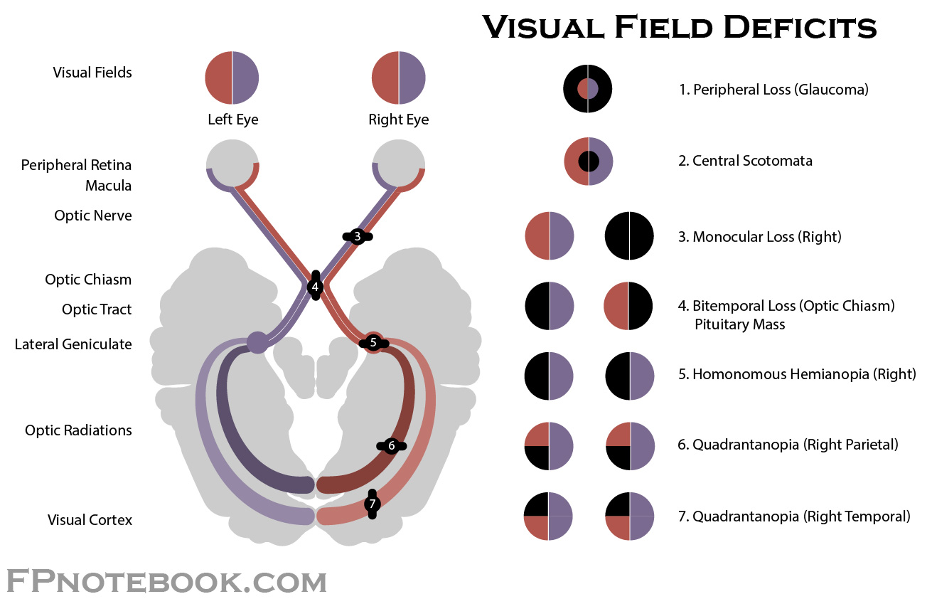
Visual Field Deficit
-
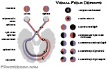
- Monocular Blindness
-
Homonymous Hemianopia (field cut affects both eyes in same region)
- Occipital lesion
-
Bitemporal Hemianopia
- Bilateral peripheral Vision Loss suggests Optic Chiasm lesion
-
-
Pupil abnormality
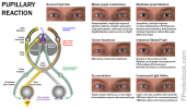
- Mid-dilated non-reactive pupil
- Afferent Pupillary Defect (sluggish or absent pupil response to light)
- Optic Nerve lesion
- Retinal lesion
-
Funduscopic Exam
-
Retinal Detachment
- Affected Retina will have the pale billowing appearance of a parachute
- In non-dilated Eye Exam, Ocular Ultrasound has better sensitivity
- Red Reflex absent
- Cherry red spot (red Macula)
- Retinal Hemorrhage
- Optic disc swelling
-
Retinal Detachment

VIII. Management
- Rapid assessment and management if acute CNS event is suspected
- Indications for emergent referral to ophthalmology
- Keratitis
- Endophthalmitis
- Retinal Detachment
- Retinal Hemorrhage or Vitreous Hemorrhage
- Optic Neuritis
- Occipital infarction
- Central Retinal Artery Occlusion
- Acute angle closure Glaucoma
- Ischemic Optic Neuropathy
- Conditions with specific immediate temporizing measures by emergency provider
- Central Retinal Artery Occlusion
- Acute angle closure Glaucoma
- Ischemic Optic Neuropathy
IX. References
- Hartmann (2016) Crit Dec Emerg Med 30(6): 3-11
- Trobe (2012) Physician Guide to Eye Care, p. 31-35