
II. Definitions
- Occipital Lobe
- Occipital Lobe primarily functions to receive and process visual input
- Brodmann Area 17 (V1) receives sensory input from the lateral geniculate nucleus in the Thalamus
- Subsequent signal processing
- Ventral stream (V2, Area 18) for form recognition and object representation
- Dorsal stream which processes motion and spatial orientation
- Further visual processing is performed in V4, a part of the Associative visual cortex (Area 19).
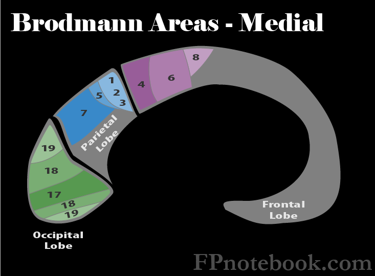
III. Anatomy: Brodmann Areas of Occipital Lobe
- Images
- Primary visual cortex (V1, Area 17)
- Receives sensory input from the lateral geniculate nucleus in the Thalamus.
- Lesions to this Primary visual cortex result in blindness of the contralateral Visual Field.
- Signals are then passed to the ventral stream (V2)
- Form recognition and object representation
- Signals are passed to the dorsal stream
- Processes motion and spatial orientation
- Secondary Visual Cortex (V2, Area 18)
- Part of the ventral stream, V2 receives visual input from the Primary visual cortex (V1)
- Processes the information for form recognition and object representation
- Also sends signals to the Associative visual cortex (V4) for further processing
- Associative visual cortex (V3, V4, V5, Area 19)
- Part of the ventral stream, V4 receives visual input from the Secondary Visual Cortex (V2)
- Further processes the information for form recognition and object representation
- Lesions to V4 will result in visual Agnosia (difficulty in object recognition)

IV. Exam
- Matches colors and objects (if unable to name them)
V. Signs: Brain Lesions
- Contralateral Hemianopia