
II. Background
- Vision is among the most complex neurologic functions of the human body
- Eye is a high-end camera
- Mechanisms to adjust aperture (pupil size) and focal length (lens shape)
- Projected onto a more than 500 Megapixel Retina
- Contains over 120 Million rods (black and white) and 6 Million cones (color Vision)
- One third of the Cranial Nerves (CN 2, 3, 4, 6) are dedicated to Vision, Extraocular Movement and pupillary reflex
- Most of the Occipital Lobe is dedicated to the processing of visual input
III. Anatomy: Visual Pathway
- Background
- Visual Field Overview
- Objects in the Visual Field are projected onto each eye's Retina as a mirror image
- Upper right Visual Field localizes to the lower left Retina of each eye
- Signals from each Retina follow the Optic Nerve to the Optic Chiasm, in the region of the Pituitary Gland
- Left Visual Field signals from each Retina (left medial and right lateral) join at Optic Chiasm
- Left Visual Field signals follow the Optic Nerve to the right Lateral Geniculate Body
- Right Lateral Geniculate Body signals follow right Optic Radiations
- Upper Visual Fields through the Temporal Lobe
- Lower Visual Fields through the Parietal Lobe)
- Signals terminate in the right Occipital Lobe visual cortex (Brodmann Area 17-19)
- Right Visual Fields are similarly routed to the left Occipital Lobe
- Visual Field Overview
- Light Pathway
- Light is refracted from Cornea and lens through pupil onto the Retina
- Light Refraction: Human eye refracts light at two locations
- See Refractive Error
- Cornea (66% of eye's focusing power)
- Fixed focusing power
- Non-Refractive Errors (e.g. Cataracts, Corneal opacities) may disrupt light pathway
- Crystalline lens (33% of eye's focusing power)
- Accommodation changes lens shape to focus objects
- Pupil is analogous to a camera aperture
- Shutter aperture decreases in size under bright light conditions and for sharper focus
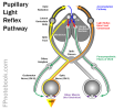
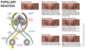
- Pupil constricts under bright light and with near focus (e.g. reading) known as accomodation
-
Retina (each eye)
- Components
- Pathway
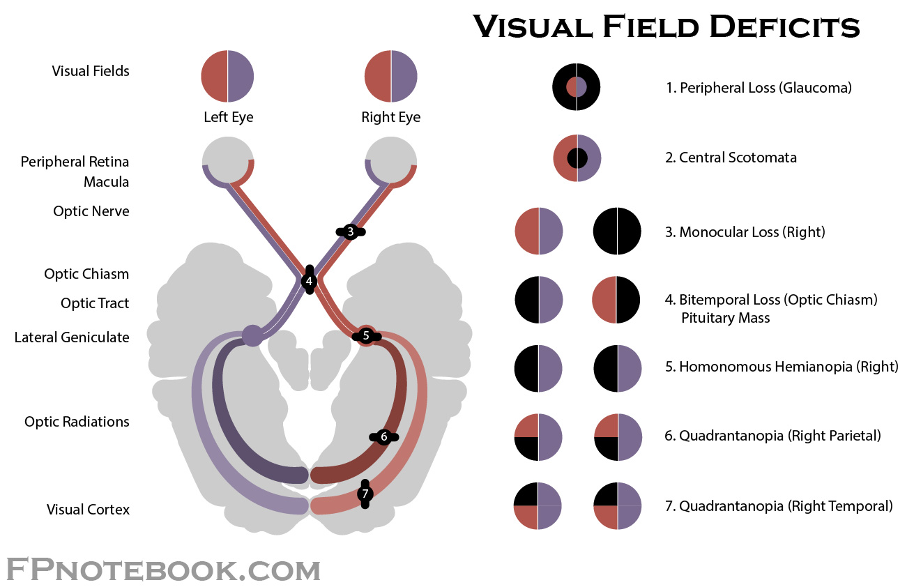
- Visual Field Deficits
- Scotomata
- Glaucoma (peripheral Vision Loss)
-
Optic Nerve (each eye)
- Components
- Pathway
- Optic Nerve fibers from the Retina lateral to the fovea course to the ipsilateral brain
- Optic Nerve fibers from the Retina medial to the fovea course to the contralateral brain
- Visual Field Deficits
- Complete loss of Vision from affected eye
- Example: Left Optic Nerve lesion results in complete blindness of the left eye, but the right eye is unaffected
- Optic Chiasm (central, near pituitary)
- Components
- Left Retinal fibers (right Visual Field) join from each eye, forming Optic Tract to left Cerebral Hemisphere
- Right Retinal fibers (left Visual Field) join from each eye, forming Optic Tract to right Cerebral Hemisphere
- Visual Field Deficits
- Bilateral loss of lateral Visual Fields
- Example: The left eye cannot see the left Visual Field and the right eye cannot see the right Visual Field
- Components
- Optic Tract (each hemisphere)
- Components
- Left Optic Tract
- Right Optic Tract
- Visual Field Deficits
- Vision Loss in each eye of the contralateral Visual Field
- Example: Left Optic Tract lesion results in a right sided Visual Field cut for each eye
- Components
- Lateral Geniculate Body (each hemisphere)
- Optic Radiation (each hemisphere)
- Components
- Parietal Lobe Optic Radiation
- Transmits superior Retina signals to Occipital Lobe above the calcarine fissure
- Temporal Lobe Optic Radiation
- Transmits inferior Retina signals to Occipital Lobe below the calcarine fissure
- Parietal Lobe Optic Radiation
- Visual Field Deficits
- Affected Optic Radiation results in a quarter segment Vision Loss
- Example: Left Parietal Optic Radiation defect results in a right upper Visual Field cut
- Components
-
Occipital Lobe (each hemisphere)
- Components
- Foveal (central Retina) is represented in the posterior Occipital Lobe
- Peripheral Retina is represented in the more anterior Occipital Lobe
- Visual Field Deficits
- Affected Occipital Lobe results in a quarter segment Vision Loss
- Central Vision (posterior Occipital Lobe)
- Peripheral Vision (anterior Occipital Lobe)
- Example: Left posterior occiput lesion above the calcarine fissure
- Results in a right upper, central Visual Field cut
- Affected Occipital Lobe results in a quarter segment Vision Loss
- Components
IV. Anatomy: Images
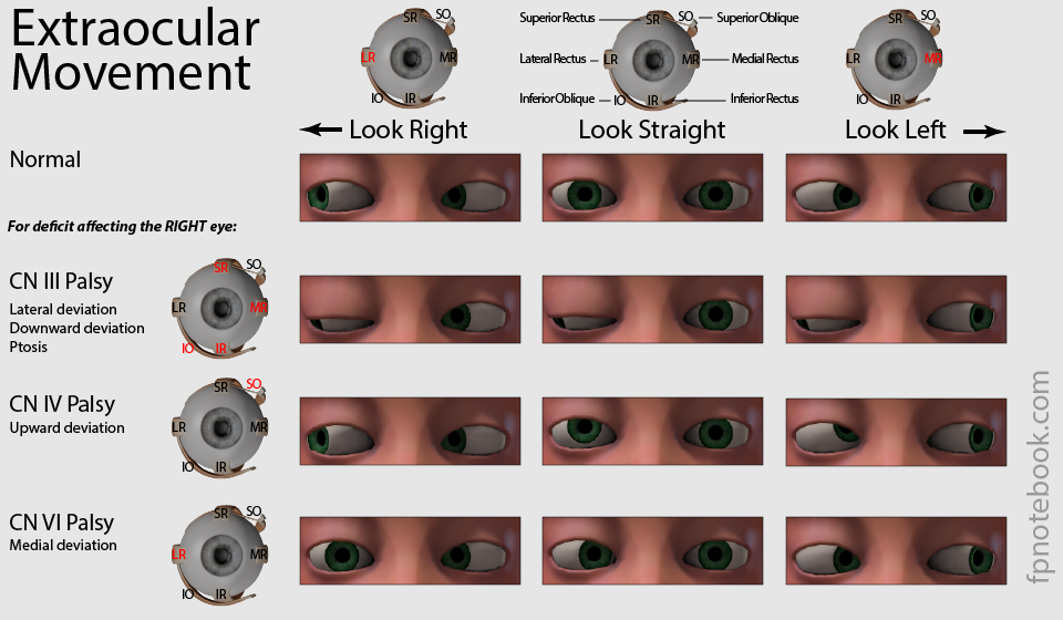
- See Extraocular Movement
- See Retinal Anatomy
- Nerves
 Lewis (1918) Gray's Anatomy 20th ed (in public domain at Yahoo or BartleBy)
 Lewis (1918) Gray's Anatomy 20th ed (in public domain at Yahoo or BartleBy)
 Lewis (1918) Gray's Anatomy 20th ed (in public domain at Yahoo or BartleBy)
 Lewis (1918) Gray's Anatomy 20th ed (in public domain at Yahoo or BartleBy)
 Lewis (1918) Gray's Anatomy 20th ed (in public domain at Yahoo or BartleBy)
 Lewis (1918) Gray's Anatomy 20th ed (in public domain at Yahoo or BartleBy)
 Lewis (1918) Gray's Anatomy 20th ed (in public domain at Yahoo or BartleBy)
 Lewis (1918) Gray's Anatomy 20th ed (in public domain at Yahoo or BartleBy)


V. References
- Goldberg (2014) Clinical Neuroanatomy, Medmaster, p. 40-54
- Netter (1997) Atlas Human Anatomy, ICON Learning, p. 114, 126