
II. Characteristics
- Monocytes form in Bone Marrow from Myeloblasts as with other Granulocytes (Basophils, Eosinophils, Neutrophils)
- Monocytes move from peripheral blood into tissue and become Macrophages
- Transformation to Macrophages controlled by Cytokines
- Functions
- Bacteriocidal activity without Phagocytosis
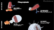
- Phagocytosis of dead or damaged cells and Bacteria (similar to Neutrophils)
- Macrophages directly perform the Phagocytosis
- Resulting Phagosome fuses with Lysosomes to digest the ingested material within the Phagosome
- Granuloma formation (epitheliod cells, multinucleated giant cells)
- Walling off of infection in response to chronic inflammation
- Result when Macrophages cannot lyse phagocytized material (e.g. silica, Asbestos, immune complexes)
- Macrophages expand, increasing cytoplasm, to form epitheliod cells
- Macrophages fuse to form multinucleated giant cells
- Monocyte Morphology on Blood Smear
- Mononuclear Leukocyte (same class as Lymphocyte)
- Slightly larger than a Lymphocyte
- Kidney shaped nucleus
- Macrophages (and related phagocytic cells) have specific names in certain tissues
- Alveolar Macrophage (lung)
- Dendritic Cells (Spleen, Lymph)
- Kuppfer Cell (Liver)
- Langerhans Cell (Skin)
- Dendritic Cell precursors
- Acute inflammation results in Langerhans Cell transit to regional Lymph Nodes
- Langerhans Cells transition to Dendritic Cells, carrying Antigen to present to Lymphocytes
- Mesangial Cell (Kidney)
- Microglia (Central Nervous System)
- Osteoclast (Bone)

III. Labs: Normal
- Range: 2-8% of White Blood Cells
IV. Causes: Increased
- Infection
- Inflammatory Bowel Disease
- Sarcoidosis
- Neoplasms
- Monocytic Leukemia
- Lymphoma
- Multiple Myeloma
V. Causes: Decreased
- Aplastic Anemia
- Lymphocytic Anemia
- Glucocorticoids
VI. Evaluation: Monocytosis (Monocytes >880/mm3)
- See Leukocytosis
- History and potential causes
- Travel and contagious contacts
- Diagnostics (consider)
- Chest XRay
- Specific infection testing (e.g. Monospot, PPD or quantiferon)
- Acute phase reactants (ESR, CRP)
VII. References
- Goldberg (2014) Clinical Physiology, Medmaster, Miami, p. 68-9
- Saiki in Friedman (1991) Medical Diagnosis, p. 227
- Abramson (2000) Am Fam Physician 62(9):2053-60 [PubMed]
- Riley (2015) Am Fam Physician 92(11):1004-11 [PubMed]