
II. Definitions
- Cardiac Output (CO)
- Quantity of blood pumped per minute through the aorta and into the peripheral circulation
- Cardiac Output is proportional to (arterialPressure / totalPeripheralResistance)
- CO = SV * HR
- Where SV = Stroke Volume
- Where HR = Heart Rate
- Normal Adult
- Range: 4-8 L/min (typical 70 kg male = 5.25 L/min)
- Cardiac Index (CI)
- Average stroke output of heart per minute (Cardiac Output) adjusted for body surface area
- CI=CO/BSA
- Where CO = Cardiac Output
- Where BSA = Body Surface Area
- Normal Adult
- Range: 2.4 - 4 L/min/m2 (60% of csardiac output in the normal, average sized adult)
- Stroke Volume (SV)
- Stroke Index (SI)
- Average stroke output of heart per cycle (Stroke Volume) adjusted for body surface area
- SI = CI/HR
- Where CI = Stroke Index
- Where HR = Heart Rate
- Normal Adult
- Range: 20-40 ml/m2
- Ejection Fraction (EF)
- Reflects the percentage of blood ejected from the ventricle
- EF = SV/EDV
- Where SV = Stroke Volume
- Where EDV = End-diastolic volume
- Normal Adult
- Left Ventricular Ejection Fraction (LVEF): 55-70%
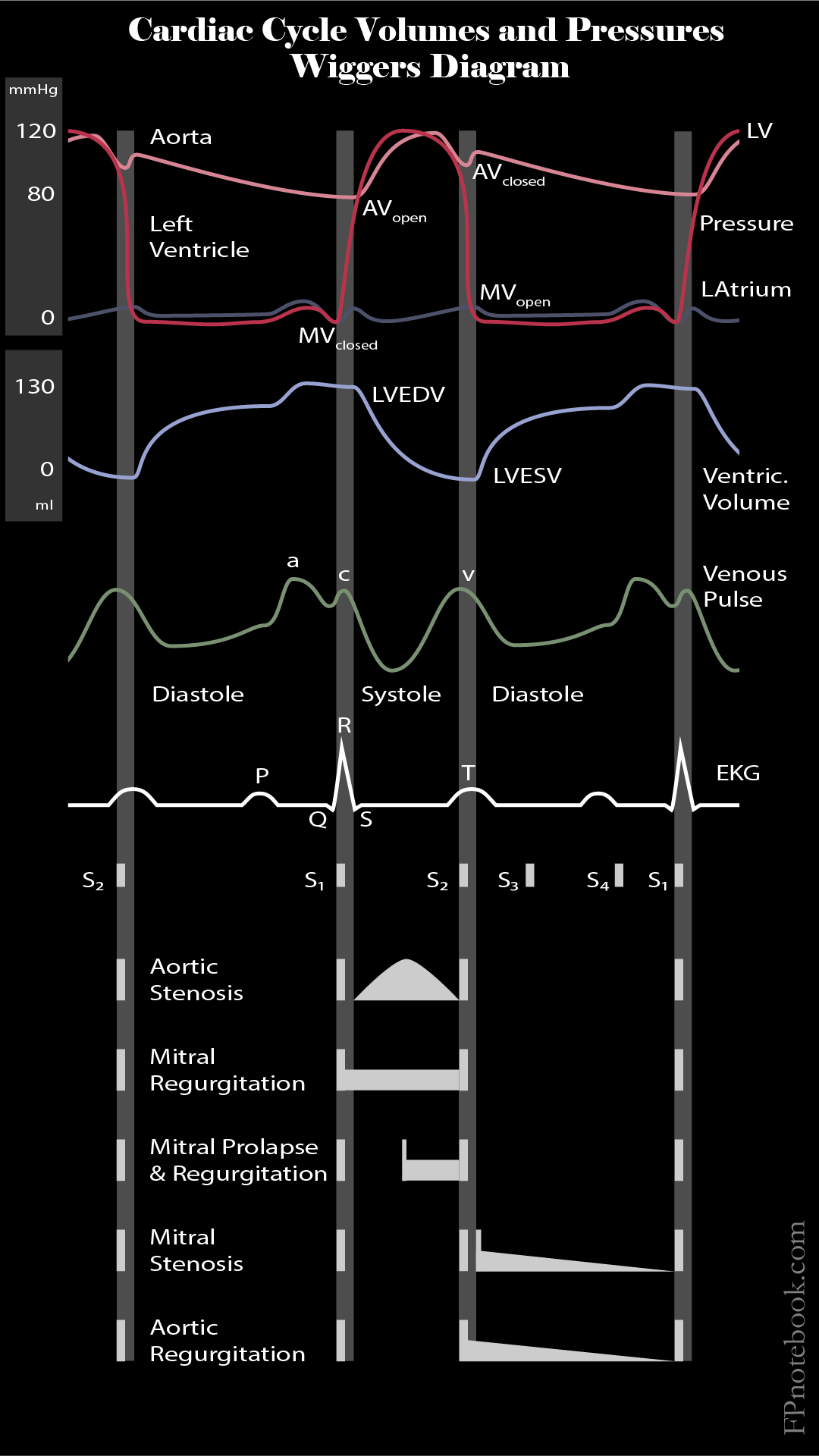
III. Images
-
Cardiac Cycle Volumes and Pressures (Wiggers Diagram)
IV. Physiology
- Stroke Volume components
- Ventricular Preload (based on venous return of Circulatory Volume and ventricular compliance)
- End-diastolic volume (EDV)
- Increases Cardiac Output in the normal heart
- Results in Fluid Overload and third spacing in Congestive Heart Failure
- End-systolic volume (ESV) depends on ejection fraction (percentage of EDV ejected from left ventricle)
- Ventricular Contractality
- Force of ventricular contraction or inotropy
- Increased contractility increases Stroke Volume and Cardiac Output
- Ventricular Afterload (systemic Blood Pressure)
- Force against which the left ventricle pumps
- Increased Afterload decreases Stroke Volume and Cardiac Output
- Ventricular Contractality
- Ventricular Preload (based on venous return of Circulatory Volume and ventricular compliance)
- Cardiac Output components
- Product of Heart Rate and Stroke Volume
- Heart Rate is the first compensatory mechanism to increase Oxygen Delivery to the tissues
- Heart is most efficient with lower Heart Rate and higher Stroke Volume
- Myocardial Oxygen Demand
- Oxygen requirements increase with venticular Muscle wall tension
- Ventricular Muscle wall tension is related to Ventricular Preload and Ventricular Afterload
- Ventricular Preload is the increasing ventricular wall tension that increases with ventricular filling
- Ventricular Afterload is the constant tension in ventricular Muscle as it contracts and shortens
- Wall tension increases with ventricular volume (ventricular radius) and ventricular pressure (Afterload)
- Laplace Equation = Tension (T) = Pr
- where P = ventricular pressure (Ventricular Afterload)
- where r = ventricular radius
- Mediators of Cardiac Output
- Positive Inotropes increase contractility
- Positive Chronotropes increase Heart Rate
- Positive Dromotropes increase cardiac conduction velocity
V. Diagnostics: Clinical Assessment of Cardiac Output (and SV, Preload, Afterload and Contractility)
-
Heart Rate and rhythm
- Palpated pulse
- Pulse Oximetry
- Telemetry or Electrocardiogram
- Right Ventricular Preload
- Left Ventricular Preload
- Symptoms (Orthopnea or Dyspnea on exertion)
- Signs (Pulmonary Edema, rales)
- Imaging (Lung Ultrasound)
- Right Ventricular Afterload
- Mean pulmonary artery pressure or wedge pressure
- Left Ventricular Afterload
- Mean arterial pressure
- Cardiac Contractility
- Echocardiogram (ejection fraction, Stroke Volume)
- Cardiac Output
- Pulmonary artery catheter
- Arterial catheter (arterial waveform analysis)
- Esophageal doppler (descending aorta flow)
VI. Resources
- Wikipedia Stroke Volume
VII. References
- Goldberg (2014) Clinical Physiology, Medmaster, Miami, p. 49-50
- Killu and Sarani (2016) Fundamental Critical Care Support, p. 93-114
- Marino (2014) ICU Book, 4th Ed Wolters-Kluwer p. 144-6