
II. Definitions
- Central Venous Pressure (CVP)
- Grouping of venous pressures that are equivalent to one another
- Superior Vena Cava Pressure
- Right Atrial Pressure (RAP)
- CVP is also equivalent to the right ventricular end diastolic pressure (RVEDP) and Preload
- Only if no tricuspid insufficiency
- Grouping of venous pressures that are equivalent to one another
- Right Ventricular End-Diastolic Pressure (RVEDP)
- Equivalent to CVP (in the absence of tricuspid insufficiency)
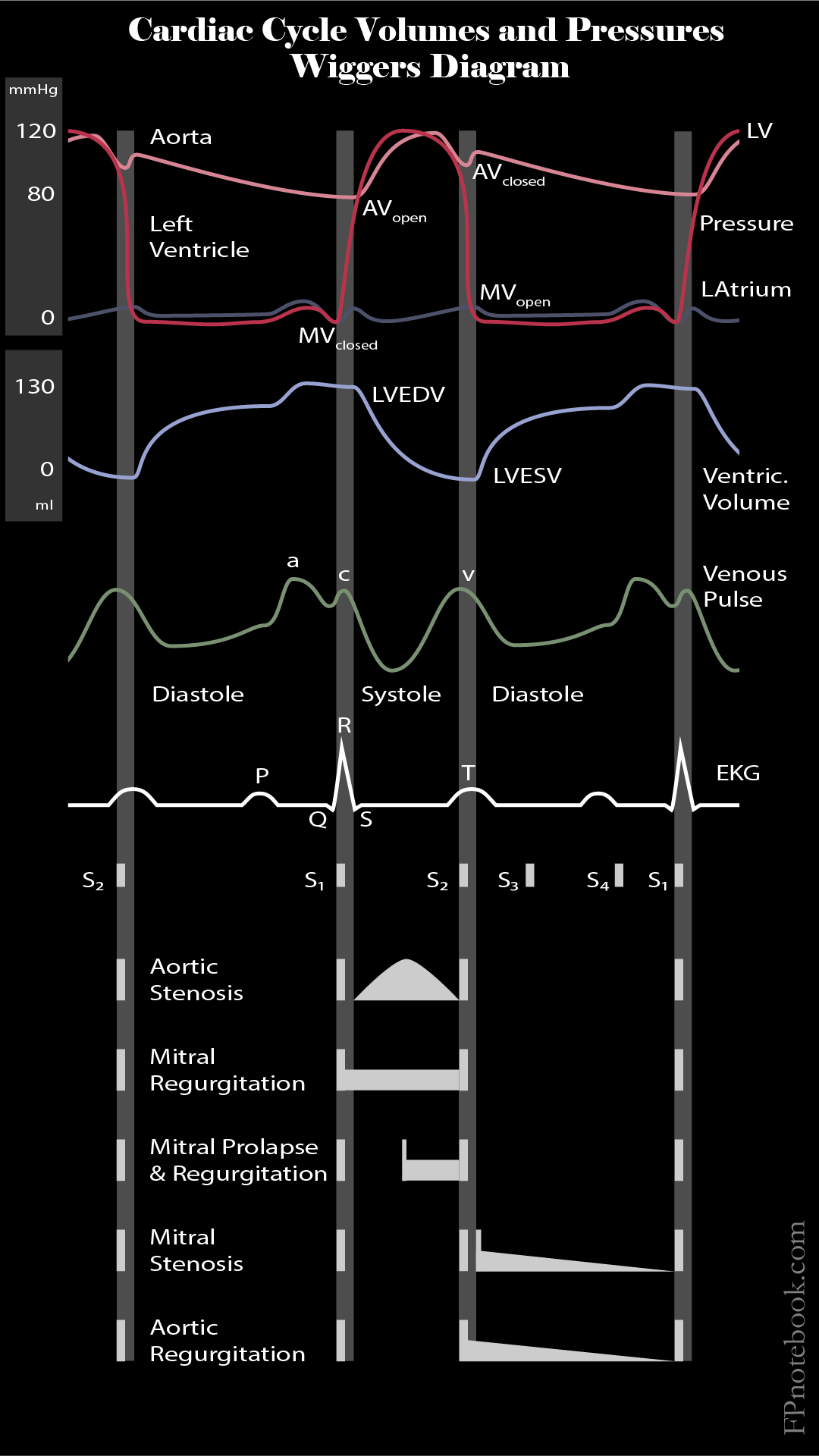
III. Images
-
Cardiac Cycle Volumes and Pressures (Wiggers Diagram)
IV. Technique: Central Venous Pressure (CVP)
- Catheters
- Central venous catheter or
- Pulmonary artery catheter (Swans-Ganz Catheter) via proximal port in atrium
- Contrast with distal port in pulmonary vessels (pulmonary wedge pressure) reflecting left atrial pressure
- PICC Lines are not used for CVP measurement due to long length
- Theoretically could be used with saline infusion
- Black (2000) Crit Care Med 28:3833-36 [PubMed]
- Transducer
- Fluid-filled device measures pressure readings from catheter
- Placed at the same level as the right atrium
- Landmarks for the supine patient
- Intersection of the fourth intercostal space and the mid-axillary line
- Landmarks for the semi-recumbent patient (<60 degrees)
- Below the sternal angle by 5 cm (directly down)
- Landmarks for the supine patient
- Measurement
- Record at the end of expiration (when intrathoracic pressure is closest to atmospheric pressure)
- CVP varies with respiration (spontaneous and Mechanical Ventilation)
- CVP decreases with spontaneous inspiration (decreased intrathoracic pressure)
- CVP increases with Mechanical Ventilation (positive pressure increases intrathoracic pressure)
V. Interpretation
- Normal Central Venous Pressure (CVP or RAP, Right Atrial Pressure)
- Normal CVP 2 to 8 mmHg (some references list 0 to 5 mmHg)
- Measurements vary as much as 4 mm Hg in the same patient under the same conditions
- CVP change is only significant if changes more than 4 mmHg
- Other Normal Right Sided Pressures
- Right Ventricular Pressure = 25/2 mmHg
- Pulmonary Artery Pressure (PAP) = 25/12 mmHg
- Mean Pulmonary Artery Pressures = 16 mmHg
- Mean Pulmonary Capillary Wedge Pressure (or left atrial pressure): 9 mmHg
- CVP may be increased initially despite significant volume depletion
- Consider underlying COPD, Vasoconstriction
- Consider increased intrathoracic pressure (Tension Pneumothorax, Positive Pressure Ventilation)
- Consider Diastolic Dysfunction with decreased right ventricular compliance (MI, Sepsis, valvular dysfunction)
- Peristently increased CVP suggests adequate volume replacement or Fluid Overload
- CVP that is markedly increased
- Fluid Overload (Edematous States such as Congestive Heart Failure, liver failure, Renal Failure)
- Cardiac Tamponade
- Tension Pneumothorax
- Central catheter malposition
- CVP that is low with signs of shock and minimally rises with fluid bolus
- Continue Fluid Replacement (or Blood Products in the case of Hemorrhagic Shock)
- Consider source of ongoing losses
- CVP that is decreasing with signs of shock
- Aggressively search for ongoing losses (e.g. Hemorrhagic Shock)
- Continue Fluid Replacement (or Blood Products in the case of Hemorrhagic Shock)
- Vasopressors as needed
VI. Precautions
- CVP is an indirect, invasive and often inaccurate surrogate for Ventricular Preload and volume status
- Consider Inferior Vena Cava Ultrasound for Volume Status as an alternative
- End diastolic pressure (e.g. CVP and Wedge Pressure) correlates poorly with end diastolic volume even in healthy patients
- Hence CVP and Wedge Pressure are unreliable markers of ventricular filling and volume status
- CVP is falsely elevated with increased intrathoracic pressure (e.g. PPV, PEEP)
VII. References
- Marino (2014) ICU Book, 4th Ed Wolters-Kluwer p. 157-9
- (2012) ATLS Manual, 9th ed, American College of Surgeons
- Killu and Sarani (2016) Fundamental Critical Care Support, p. 93-114