
II. Exam: Telemedicine
- See Telemedicine
- Perform observation (see below) and compare with opposite side
- Perform Wrist Range of Motion as below
- Patient palpates their own wrist for point of maximal tenderness
- Patient may perform specific testing with coaching
- Carpal Tunnel Syndrome (Phalen's Test, Tinel's Test)
- De Quervain's Tenosynovitis (Finkelstein Test)
III. Exam: Observation (comparing with opposite side)
- Erythema
- Deformity
- Swelling or joint effusion
- Ecchymosis (recent Trauma)
- Overlying skin changes
- Scars suggesting old Trauma
IV. Exam: Normal Range of Motion
V. Exam: Triangular Fibrocartilage Complex (TFCC)
- Locate depression between Pisiform and ulnar styloid
- Tenderness distal to this point suggests TFCC Injury
- Patient and examiner shaking hands
- Patient tries to supinate or pronate wrist
- Pain or decreased ROM suggests TFCC Injury
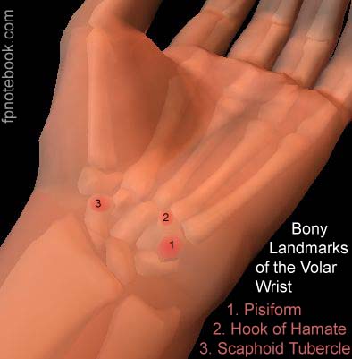
VI. Exam: Bony Landmarks on Volar Surface
- Images
-
Pisiform
- Ulnar side of palm just proximal to palmar crease
- Flexor carpi ulnaris inserts on Pisiform
- Identify by opposing thumb and fifth finger
- Assess for tenderness at bony prominence
- Hook of Hamate
- Hypothenar wrist, 1 cm distal to flexor crease
- Identification Method 1
- Start at Pisiform
- Move 1-2 finger breadths toward midline
- Identification Method 2
- Flex wrist and hook of Hamate becomes prominent
- Palmaris longus (if present) courses above Hamate
- Identify by opposing thumb and fifth finger
- Absent in 10% of patients
- Do not confuse with flexor carpi radialis
-
Scaphoid tubercle
- Palpable at extensor carpi radialis at palmar crease
- Press at rest to assess for tubercle Fracture
- Press while moving from ulnar to radial deviation
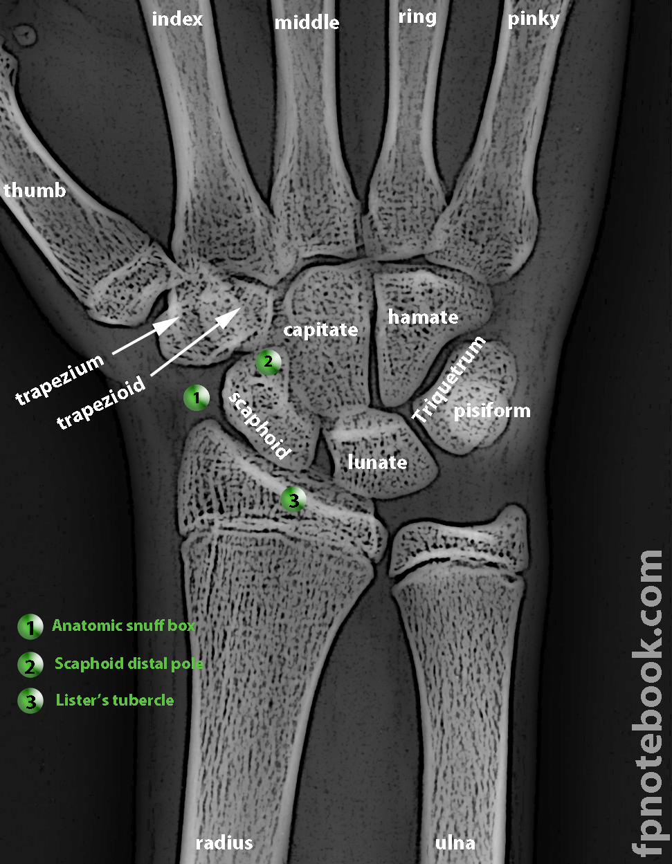
VII. Exam: Bony Landmarks on Dorsal Surface
- Images
- Anatomic Snuff box
- Radial border
- Extensor pollicis brevis
- Abductor pollicis longus
- Ulnar border
- Extensor pollicis longus
- Radial border
-
Scaphoid distal pole
- Located at anatomic snuff box
- More easily palpated with wrist in ulnar deviation
- Scaphoid is the most commonly Fractured Carpal Bone
- See Scaphoid Fracture Signs
-
Carpal Bones on radial side of wrist
- Shuck Test evaluates for inflammation/instability
- Triscaphe Joint
- Thumb follows second finger proximally over dorsum
- Thumb falls into triscaphe joint depression
- Lister's Tubercle (Radial Tubercle)
- Distal radius prominence on wrist dorsum
- Palpate radius dorsum while patient flexes wrist
- Lines up with Third Metacarpal
- Lister's Tubercle
- Lunate
- Capitate
- Third Metacarpal
- Scapholunate joint or interval
- Most common carpal dislocation
- Scaphoid Shift Test evaluates scapholunate injury
- Identification (1.5 cm distal to Lister's Tubercle)
- Examiners finger starts over Third Metacarpal
- Trace Third Metacarpal proximally over dorsum
- Examiner's finger falls into depression
- Depression represents scapholunate joint
-
Lunate
- Second to Scaphoid as most common wrist Fracture
- Identification
- Identify with patient flexing wrist
- Most prominent area on dorsum of flexed wrist
- Lunate sits on ulnar side of scapholunate joint or
- Follow Lister's tubercle distally to 3rd Metacarpal
- Identify with patient flexing wrist
VIII. Exam: Specific to Wrist Overuse syndromes
- See Overuse Syndromes of the Hand and Wrist
- Carpal Tunnel Syndrome (Phalen's Test, Tinel's Test)
- De Quervain's Tenosynovitis (Finkelstein Test)
- Intersection Syndrome (tender at dorsal distal radius)
IX. References
- Hoppenfeld (1976) Exam Spine/Extremities, p. 59-104
- Daniels (2004) Am Fam Physician 69(8):1941-48 [PubMed]
- Forman (2005) Am Fam Physician 72:1753-8 [PubMed]
- Yedlinsky (2021) Am Fam Physician 103(3):147-54 [PubMed]