
II. Indication
III. Exam: Telemedicine
- See Telemedicine
- Perform observation (see below) and compare opposite side
- Patient points to point of maximal tenderness (e.g. snuffbox)
- Patient performs finger range of motion
- Test specific Hand Neurovascular Exam (see below)
- Patient performs hand strength examination
- Make a fist and then fully extend fingers
- Patient specifically tests DIP and PIP extension and flexion against their own resistance
IV. Observation: General
-
General appearance (comparing with opposite side)
- Erythema
- Deformity
- Swelling or joint effusion
- Ecchymosis (recent Trauma)
- Overlying skin changes
- Nail changes
- Atrophy
- Scars suggesting old Trauma
-
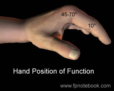
Hand Position of Function
- Inability to assume position of function is a red flag
- Position of Function Image
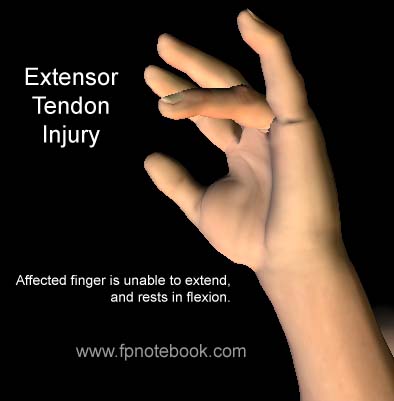
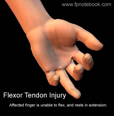
V. Observation: Fixed digital flexion or extension
- Fixed single finger extension: Flexor Tendon Injury
- Image
- Description: Observe with hand at rest
- Affected finger in complete extension
- Other fingers PIP and DIP at 10 degrees flexion
- Injuries
- Image
- Fixed single finger flexion: Extensor Tendon Injury
VI. Observation: Finger rotational deformity
VII. Observation: Skin changes
- Vascular compromise signs
- Capillary Refill >2 seconds
- Also check radial pulse and ulnar pulse
- Consider Allen Test for ulnar artery distribution
- Digital nerve injury
- Two Point Discrimination requires >5 mm apart
- Skin Color change
- Anhidrosis of finger involved
- Blanched or hyperemic Skin Color
VIII. Exam: Brief tendon evaluation
- Observe for fixed digital flexion or extension
- See above
- Extend DIP joint of affected finger
- Start with all fingers in extension
- Flex PIP joint of affected finger only
- Injury
- Flexor digitorum profundus injury (Jersey Finger)
- Testing
- Test with all fingers in extension
- Hold affected finger DIP in extension
- Patient instructed to flex affected PIP joint
- Injury
- Flex DIP joint of affected finger only
- Consider flexor digitorum superficialis injury
- Specific test for index finger
- Patient pulls a paper between both hands
- Paper held by each hand pinching paper
- Each hand uses opposed thumb and index finger
- Injury of flexor digitorum superficialis signs
- DIP hyperflexes
- PIP hyperextends
- Patient pulls a paper between both hands
- Flex PIP joint of affected finger only