
II. Epidemiology
- Mallet Finger is the most common closed finger Tendon Injury
III. Mechanism
- Forced flexion of extended distal interphalangeal joint
- Ball strikes fingertip on catching a ball
-
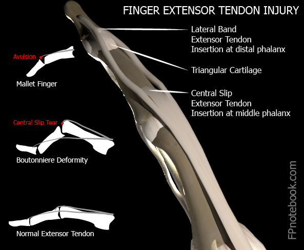
Trauma at DIP joint results:
- Avulsion of distal phalanx (Bony Mallet) as extensor tendon tears away bony insertion or
- Pure extensor tendon rupture (Tendinous Mallet)
- Tendon stretched, or partially or completely torn
- Images
IV. Symptoms
- Pain, Bruising and swelling at dorsal DIP joint
V. Signs
- DIP joint with flexion deformity at rest
- Intact flexor tendon unopposed by the ruptured extensor tendon
- Isolate the DIP joint to test active extension
- Variable loss of active finger DIP extension
- Confirm extension weakness due to extensor tendon
- Central slip at PIP joint can also extend DIP
VI. Associated Conditions
- Volar subluxation of distal phalanx with bony mallet
VII. Imaging: XRay of digit (esp. lateral)
- Assess for bony mallet at dorsal base of distal phalanx
- Type I: No avulsion fragment
- Type II: Small bony avulsion
- Type III: Avulsion with volar subluxation
- Post-reduction XRay to confirm proper alignment
- Repeat XRay every 2 weeks if bony mallet (DIP avulsion Fracture)
VIII. Management: Splinting
-
General
- Splints are equally effective: Aluminum, Stack, Ring
- Splints are as effective as surgical repair
- May participate in sports with splinted DIP
- Technique
- May use prefabricated splint instead
- Measure and cut the splint to extend from fingertip to the middle phalanx
- Should only immobilize the DIP joint (not the PIP joint)
- Smooth sharp edges
- Mold the splint to keep the DIP joint in slight hyperextension (5-10 degrees)
- Splint may be applied to either the dorsal or volar surface (author prefers volar surface)
- Tape the splint in place
- Precautions
- See Orthopedic referral indications below
- Splint should not reduce range of motion of PIP
- Splinting must be continuous for entire period (DIP must remain in extension)
- Splinting time (6-8 weeks) restarts if the finger falls back into flexion
- Delayed presentation (e.g. month old injury) requires a longer period of Splinting
- Risk of skin necrosis with Splinting
- Avoid pressure to dorsum of DIP
- Avoid hyperextension of DIP joint
- Skin will blanch if DIP hyperextended
- Assessment
- Post-reduction XRay to confirm proper alignment
- Protocol
- First 6-8 weeks
- Splint finger in neutral extension for 6-8 weeks
- Splinting must be continuous without fail
- Twenty four hours per day
- Every day for 6-8 weeks
- Hold extension when changing splint
- Support distal phalanx against flat surface
- Ask for assistance when changing splint
- Allow skin to air for 10 minutes at splint change
- Reduces maceration at splint
- Restart 8 week Splinting period if finger flexes
- Next 3-6 weeks
- Splint finger in extension only at night
- First 6-8 weeks
IX. Management: Orthopedic Referral Indications (see prognosis below)
- Joint incongruent
- Inability to passively extend DIP joint
- Suggests bone or soft tissue entrapment
- Fracture involves >30% of joint space
- Fragment displaced >2mm
- Open Growth Plate
- Bony avulsion >1/3 of distal phalanx
- Volar subluxation of distal phalanx
X. Management: Follow up
- Re-examine every two weeks until healed
- XRay every two weeks if bony avulsion
XI. Management: Anticipatory Guidance
- Warn that patient that outcome will not be perfect
XII. Prognosis
- Outcomes are similar for conservative therapy versus surgical management (regardless of referral indications above)
XIII. Complications
- Chronic loss of full distal phalanx extension
XIV. References
- Brandenburg (1996) Consultant p.331-340
- Calmbach (1996) Lecture in Minneapolis
- Dvorak (1996) Lecture in Minneapolis
- Lillegard (1996) Lecture in Minneapolis
- Warrington (2023) Crit Dec Emerg Med 37(3): 22
- Childress (2022) Am Fam Physician 105(6): 631-9 [PubMed]
- Leggit (2006) Am Fam Physician 73(5):810-6 [PubMed]
- Simpson (2001) J Hand Surg 26:32-3 [PubMed]
- Wang (2001) Am Fam Physician 63(10):1961-66 [PubMed]