
II. Images
III. Exam: Telemedicine
- See Telemedicine
- Patient should be wearing shorts (or similar) and barefoot for exam
- Camera should be positioned to adequately visualize both lower legs with adequate detail
- Perform general Ankle Exam with inspection, range of motion evaluation (see below)
- Ankle strength may be tested against gravity or against resistance (e.g. towel, resistance bands)
- Perform standing and gait exam (see below)
- Patient should palpate and point to regions of ankle or foot with maximal pain
- Include squeezing the ankle and calf (Squeeze Test, Thompson Test)
- Include proximal tibia and fibula tenderness (e.g. Maisonneuve Fracture)
- Neurovascular Exam
- Capillary Refill may be assessed if adequate lighting and camera at close position
- Dermatome distribution light Touch Sensation may be performed by patient
- Consider Ankle XRay or other imaging indications
IV. Exam: General Ankle
- Inspection (Compare both ankles without shoes or socks)
- Erythema
- Deformity
- Swelling or joint effusion
- Muscle Atrophy
- Ecchymosis (recent Trauma)
- Overlying skin changes
- Scars suggesting old Trauma
- Ankle Range of Motion (normal values)
- Ankle stability
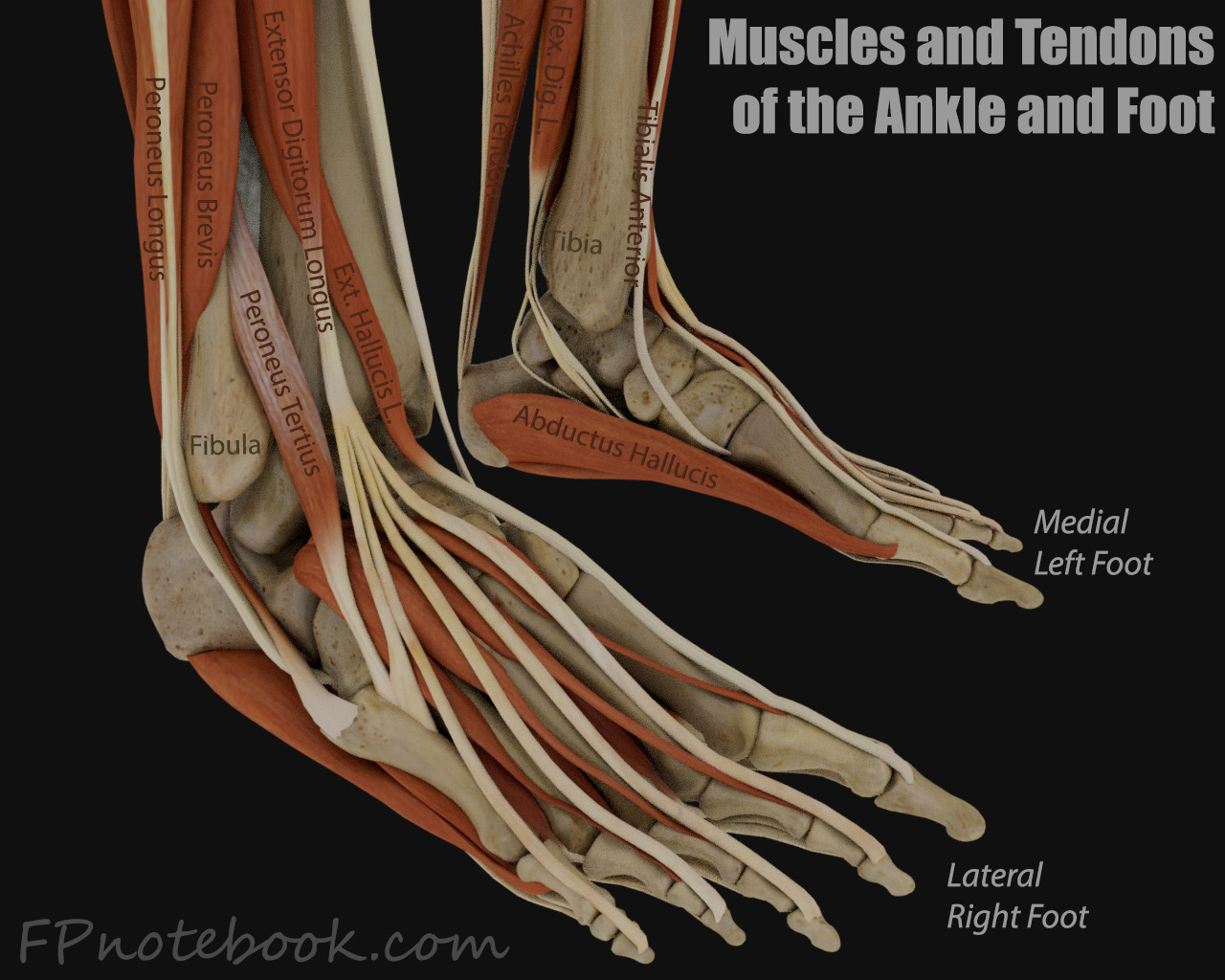
- Lateral Ankle
- Palpate the distal fibula
- Palpate the lateral ankle ligaments (torn in Lateral Ankle Sprain)
- Anterior talofibular ligament (ATFL)
- Posterior talofibular ligament (PTFL)
- Calcaneal fibular ligament (CFL)
- Medial Ankle
- Palpate the distal tibia and medial malleolus (Ottawa Ankle Rule)
- Palpate the medial deltoid ligament complex
V. Exam: Additional exam outside the ankle
- Standing Exam
- Observe foot from patient's back with feet Shoulder width apart
- Toes seen lateral to foot >2.5 suggests out toeing or hyperpronation
- Observe foot from side
- Pes cavus
- Pes Planus
- Observe gait
- Observe foot from patient's back with feet Shoulder width apart
-
Foot vascular exam
- Posterior tibial pulse
- Dorsalis pedis pulse
- Distal foot Capillary Refill
-
Foot and ankle Neurologic Exam
- Active ankle plantar flexion, ankle dorsiflexion and great toe dorsiflexion
- Toe movement
-
Foot
- Palpate the Tarsal Navicular Bone for Fracture
- Palpate the proximal fifth Metatarsal Bone (site of avulsion from peroneus brevis tendon)
- Palpate the midfoot dorsum (lisfranc joint injury)
- Leg
- Palpate the proximal fibula (for Maisonneuve Fracture)
- Thompson Test (evaluate for Achilles Tendon Rupture)
- Syndesmotic Sprain testing