
II. Anatomy
- See Pupil Constriction (Miosis) for Neuroanatomy of Pupil Constriction
- Variable sized circular hole in pigmented iris
- Images
- Pupil "Hole" or aperture size controlled by 2 Muscles
- Sphincter pupillae
- Circular Muscle constricts pupil (Miosis)
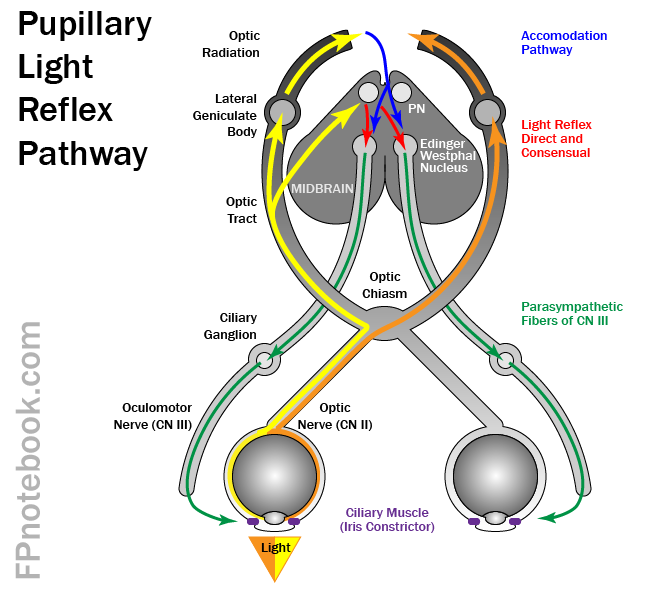
- Parasympathetic fibers on CN3 from Edinger-Westphal Nucleus innervate the ciliary Ganglion
- Dilator pupillae
- Radial Muscle dilates pupil (Mydriasis)
- Sympathetic inputs via:
- Hypothalamus
- Ciliospinal center of Budge and Waller
- Sphincter pupillae

III. Physiology
- Pupil is analogous to a camera aperture
- Shutter aperture decreases in size under bright light conditions and for sharper focus
- Pupil constricts under bright light and with near focus (e.g. reading) known as accomodation
-
Optic Nerve (CN 2)
- Transmits visual signals not only to the visual cortex
- Transmits signals via a reflex loop to the pretectal nucleus
- Signals then travel onto the Edinger-Westphal Nucleus on each side of the Midbrain
- Shining light in one eye will result in a signal to both Edinger-Westphal Nucleii
- Signals are then transmitted to the ciliary Ganglion and along the course of Oculomotor Nerve
-
Oculomotor Nerve (CN 3)
- Innervates ciliary Muscle (iris constrictor), resulting in Pupil Constriction
- Accomodation (near Vision) also results in signals to the Edinger-Westphal Nucleus
- However those signals arise from the visual cortex
- Syphilitic Pupil (Argyll-Robertson Pupil, Prostitute's Pupil)
- Affected pupil does not react
- Lesion in the Midbrain blocks signals from the pretecal nucleus
- Syphilitic Pupil does accomodate
- Signals follow a different pathway from the visual cortex
- Affected pupil does not react
- Brainstem Herniation
- . Oculomotor Nerve (CN 3) is easily compressed
- Results in a dilated pupil that does not react (Blown Pupil)
- Parasympathetic fibers from the Edinger-Westphal Nucleus to the ciliary Muscle
- Found on the outside of the Oculomotor Nerve (CN 3),
- Parasympathetic fibers are first to be injured when CN 3 is compressed
IV. Exam
- See Pupillary Light Reflex
- See Miosis (Pupil Constriction)
- See Mydriasis (Pupil Dilation)
V. Findings
- Pupil Normal in
- Pupil size changes
- Pupil distortion
- Posterior synechiae
- Inflammatory adhesion between lens and iris
- Tear drop-shaped pupil
- Suggests Globe Rupture with iris prolapse
- Posterior synechiae
- Pupil fixed, mid-dilated, slightly irregular
- Acute Glaucoma
- Absent Pupillary Light Reflex
- Structural lesion (esp. Optic Nerve)
- Hypothermia
- Barbiturate or Opiate Overdose
- Mydriatric (Eye Dilating Drop)
- Posterior synechiae
- Hippus (minimal change in light reflex seen in younger patients)
-
Relative Afferent Pupillary Defect (RAFD, unilateral with Swinging Flashlight Test)
- Ischemic Optic Neuritis
- Optic Neuritis
- Optic Nerve compression or Trauma
- Glaucoma (asymmetric)
- Optic Neuropathy (e.g. infiltrative, radiation, infectious)
- Broadway (2012) Community Eye Health 25(79-80):58-9 +PMID: 23520419 [PubMed]