
II. Anatomy
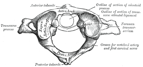
- Axis (C1)
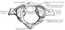 Lewis (1918) Gray's Anatomy 20th ed (in public domain at Yahoo or BartleBy)
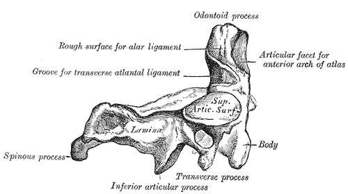
- Odontoid (C2) Lateral
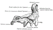 Lewis (1918) Gray's Anatomy 20th ed (in public domain at Yahoo or BartleBy)
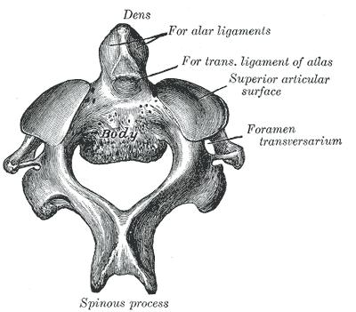
- Odontoid (C2) from Above
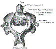 Lewis (1918) Gray's Anatomy 20th ed (in public domain at Yahoo or BartleBy)
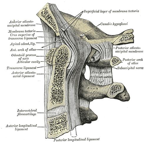
- C1 on C2
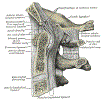 Lewis (1918) Gray's Anatomy 20th ed (in public domain at Yahoo or BartleBy)
III. Evaluation: Adequate film criteria
- C1-2 Fractures are very high risk
- More common at the extremes of age (very young and old)
- Lateral masses of C1 seen
- Entire odontoid and the superior aspect of C2 Vertebral body visualized
- Foramen magnum view (tip shot)
- XRay directed at the C1-2 interface from below the angle of the jaw
- Consider if unable to view odontoid tip on standard odontoid view
- However, can miss odontoid Fractures due to overlap (Cervical Spine CT is far preferred)
IV. Evaluation: Alignment
- Occipital condyle articulates with C1 lateral masses
- C2 Odontoid sits evenly between the C1 lateral masses (atlantodental interval)
- Spacing is symmetric to either side of the odontoid
- Asymmetry of spacing >1 mm is abnormal
- Lateral borders of c1 and c2 Vertebrae should line up
- Splaying apart (separation) of the lateral masses is abnormal
- Results in the malalignment of the C1 on C2 lateral masses
- Vertebral landmarks shifted in on one side
- Should be shifted out on other side
- Splaying apart (separation) of the lateral masses is abnormal
V. Evaluation: Bony Margins
- Odontoid should have uninterrupted cortex to C2
- Odontoid view C1 and C2 should line up
VI. References
- Tubbs and Janicki (2025) Mastering Emergency Imaging, CCME, accessed 8/7/2026