
II. Indications
III. Evaluation: General
- Mnemonic: ABCDS
- Adequate film and Alignment
- Bony landmarks
- Cartilaginous space and curvature
- Discs
- Soft tissue spaces
- Anatomy
- Odontoid (C2) Lateral
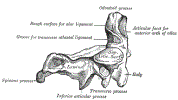 Lewis (1918) Gray's Anatomy 20th ed (in public domain at Yahoo or BartleBy)
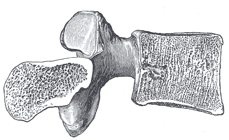
- Vertebra Lateral
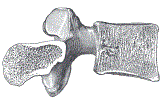 Lewis (1918) Gray's Anatomy 20th ed (in public domain at Yahoo or BartleBy)
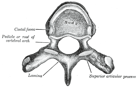
- Vertebra from above
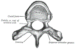 Lewis (1918) Gray's Anatomy 20th ed (in public domain at Yahoo or BartleBy)
- Odontoid (C2) Lateral
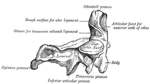
IV. Evaluation: Adequate film criteria
- Visualize all seven Vertebrae (including C7-T1)
- Major pitfall: Inadequate visualization of the entire Cervical Spine from C1 to the top of T1
- C7-T1 is a common site of Cervical Spine Injury
- Visualization of the C7-T1 interface is critical
- Maneuvers to enhance view of lower C-Spine C7-T1 (pulling Shoulders down and out of the way)
- Pull down on arms during cross-table lateral
- Swimmer's View
- Cervical Spine CT
V. Evaluation: Alignment
- Assessment criteria
- Landmarks should line up with <3mm discrepancy
- Curvature
- Normal lordotic curvature of the Cervical Spine
- Landmarks: 4 Aligment Lines
- Normal variants: Pseudosubluxation of C2 and C3
- Normal in children
- Related to large occiput altering c-spine positioning in the supine position (cross-table lateral)
- Seen in 20-40% of children under age 8 years
- Pseudosubluxation may also be seen in up to 14% of children at C3-4
- Swishchuk's Line can be used to confirm pseudosubluxation
- Spinolaminar line drawn from C1 to C3
- C2 spinolaminar line should fall within 1 mm of the C1-C3 line
- Normal in children
- Abnormal findings
- Subluxation of >=3 mm is abnormal
- Angulation >11 degrees is unstable
VI. Evaluation: Bony landmarks
- Visualize C1 anterior and posterior arch
- Trace odontoid outline from C2 Vertebrae for defects
- Trace the ring-like appearance of C2 Vertebral body (lateral masses)
- Trace unbroken outline of each Vertebrae
- Evaluate the alignment of the articulating facets and pedicles (immediately behind the Vertebral body)
- Facets have parallel aligment (roof shingle-like) without overlap
- Small linear space maintained between each facet
- Overlap may suggest possible facet dislocation
- Spinous process
- Each level of spinous process should parallel the adjacent superior and inferior spinous process
- Splaying out between spinous processes may suggest Fracture
VII. Evaluation: Cartilaginous Space
- Predental space (atlanto-dens interval, anterior atlanto-odontoid space)
- Atlas anterior margin (C1) to Odontoid process or dens (C2)
- Normal values
- Adult: <=3mm
- Child: <=4-5mm
VIII. Evaluation: Discs
IX. Evaluation: Soft Tissue spaces
- Normal Fat tissue stripe <3 mm
- Maximum normal width of soft tissue stripe anterior to Vertebral bodies (Mnemonic: 6 at 2 and 22 at 6)
- Normal Pre-Dens Space (between C1 anterior arch and C2 odontoid)
- Abnormally increased space is suggestive of ligamentous instability (e.g. Rheumatoid Arthritis, Downs Syndrome)
- Adult: <3 mm
- Child: <5 mm
X. References
- Ouellette and Tetreault (2015) Clinical Radiology, Medmaster, Miami, p. 42-50
- Tubbs and Janicki (2025) Mastering Emergency Imaging, CCME, accessed 8/7/2026