
II. Indications
III. Approach: Right Intercostal Oblique View (Right lower chest to RUQ)
- Transducer positioning
- Landmarks
- Conditions
- Right Hemothorax
- Posterior lateral wall bright attenuation artifact extends above diaphragm
- Black anechoic fluid (may have internal echoes if clotted blood)
- Lung tissue may be seen within black fluid, free floating
- Abnormal Continuation of the Spinous Line
- Hemothorax may be difficult to visualize on Ultrasound (in comparison with Pleural Effusion)
- However, Hemothorax provides an acoustic window to visualize spine continuation into the chest
- Spine is usually obscured by lung air and NOT visible on Ultrasound
- References
- (2025) FAST Exam, Hospital Procedures Course
- Right Hemothorax
- Images


IV. Approach: Right Coronal View (RUQ to RLQ, pericolic and inferior renal pole)
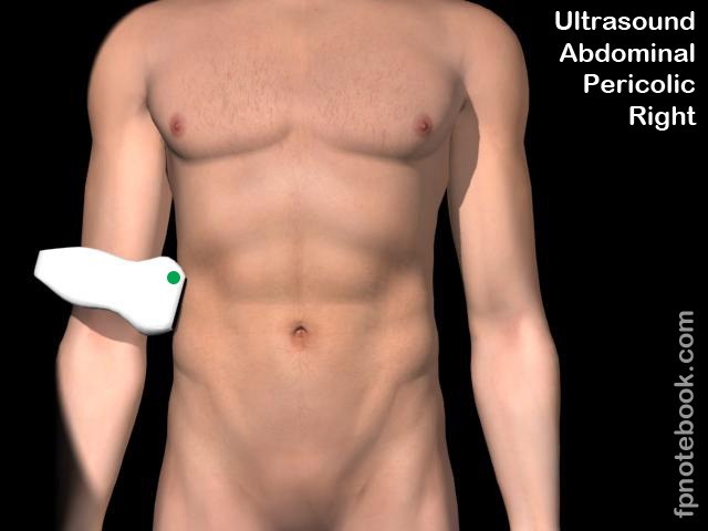
- Obtain view by tilting transducer inferiorly from right intercostal view (or dropping down 1-2 rib spaces)
- Transducer positioning
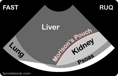
- Landmarks
- Conditions
- Blood in Morrison's Pouch (between right Kidney and liver)
- Fat may appear similar to blood
- However fat has a homogeneous speckled appearance without change in size
- Blood Clot in Morrison's Pouch is a sign of catastrophic Hemorrhage (>1 Liter of blood in Abdomen)
- Fluid in Morrison's Pouch in the absence of Trauma
- Ascites may also appear as fluid in pouch, but will be diffuse
- In association with Acute Abdomen, suggests intraabdominal catastrophe
- Fat may appear similar to blood
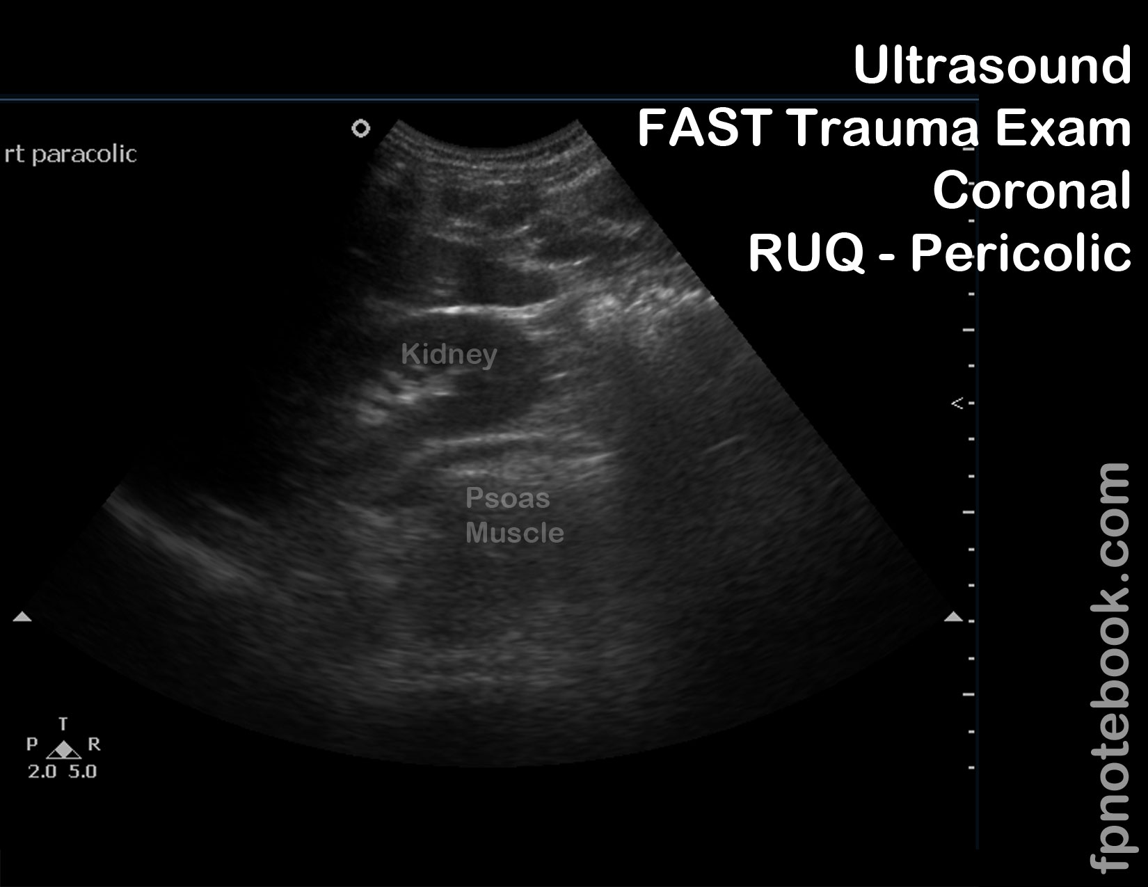
- Blood in Paracolic Gutter (anterior or superficial to right Kidney)
- In supine patients, paracolic gutter is lowest point in peritoneal cavity above pelvic brim
- Small blood accumulations may appear here first (prior to Morrison's pouch)
- Blood in Right Retroperitoneum
- Blood accumulates between Kidney and psoas Muscle
- Most blood in Retroperitoneum will be indistinguishable from Muscle
- Blood in Morrison's Pouch (between right Kidney and liver)
- Images
V. Resources
- FAST Exam RUQ (Dr. Mandavia, Sonosite)
- FAST Exam RUQ -Normal (Dr. Mandavia, Sonosite)
- FAST Exam RUQ - Hemorrhage (Dr. Mandavia, SonoSite)
VI. References
- Reardon (2016) FAST Scan, Online Video Stabroom.com, accessed 4/1/2016
- Reardon (2013) Emergency Ultrasound Course, 3rd Rock Ultrasound, Minneapolis, MN
- Alameda County Trauma Service FAST Exam
- Mateer (2012) Introduction to Trauma Ultrasound Video, GulfCoast Ultrasound, VL-95-T
- HCMC FAST Exam