
II. Precautions
- Left Pleural Effusion is often missed on FAST Exam
- Do not forget to orient probe superiorly to visualize diaphragm
- Clinically important Pleural Effusions (or Hemothorax) will be clearly seen
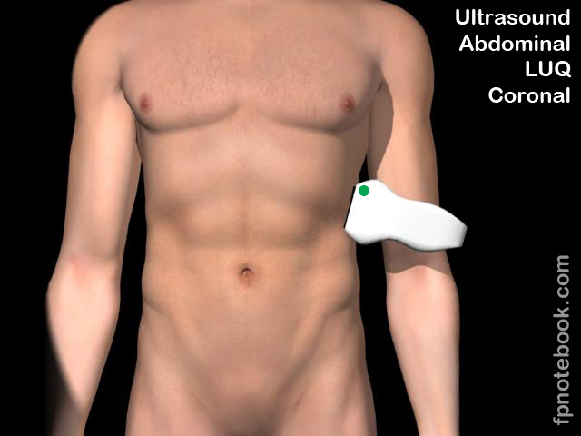
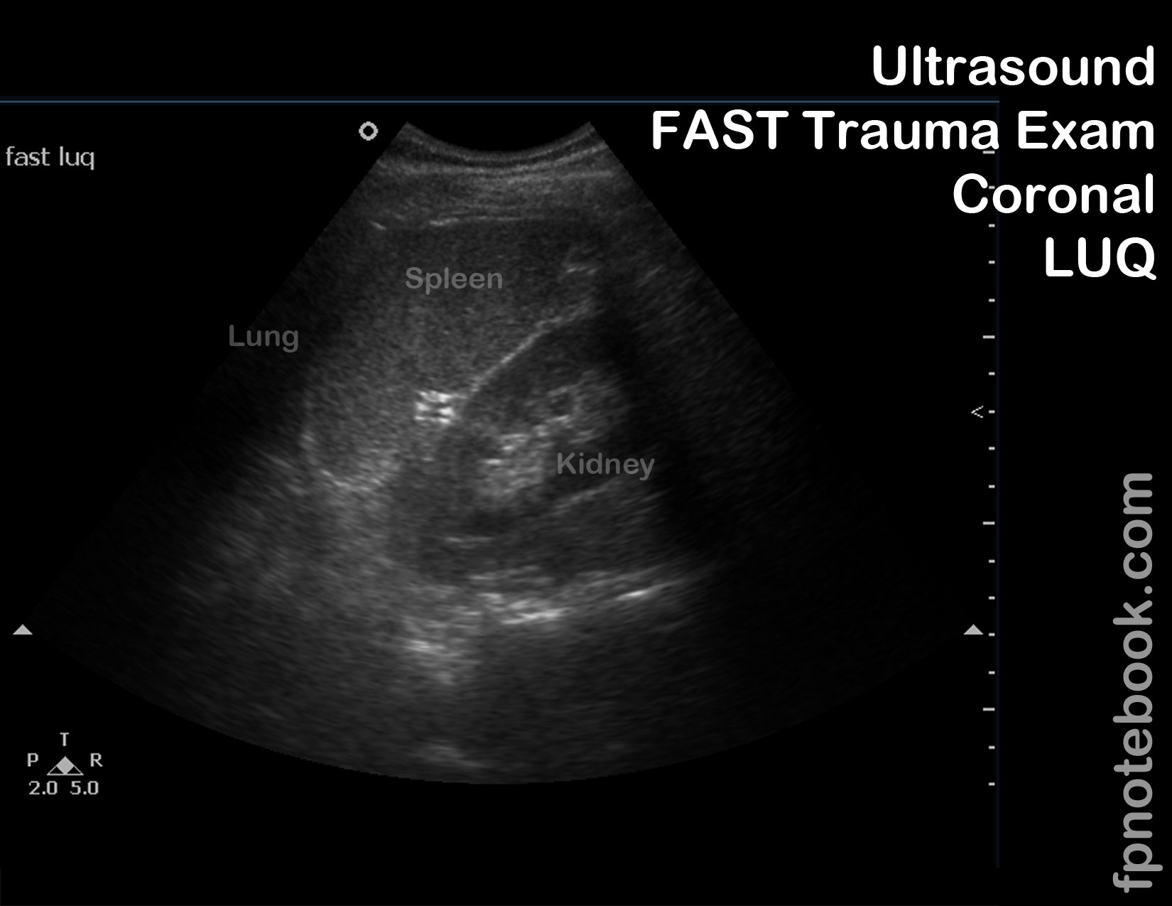
III. Approach: Left Intercostal Oblique Ultrasound View (LUQ)
- Transducer positioning
- Placement
- Hand resting on bed, holding transducer slightly above plane of bed (posterior axillary line)
- Transducer posiition is at approximately 9th intercostal space
- Axis: Long axis with indicator at 12:00
- May rotate transducer to oblique with indicator towards 1-2:00 to reduce rib shadowing
- Direction: Energy perpendicular to lateral chest towards liver
- Precautions
- Placement
- Landmarks
- Conditions
- Left Hemothorax (same findings as on right)
- Additional measures (if time to evaluate incidental findings)
- Spleen can also be measured for Splenomegaly in this view (normal <12-14 cm)
- Images
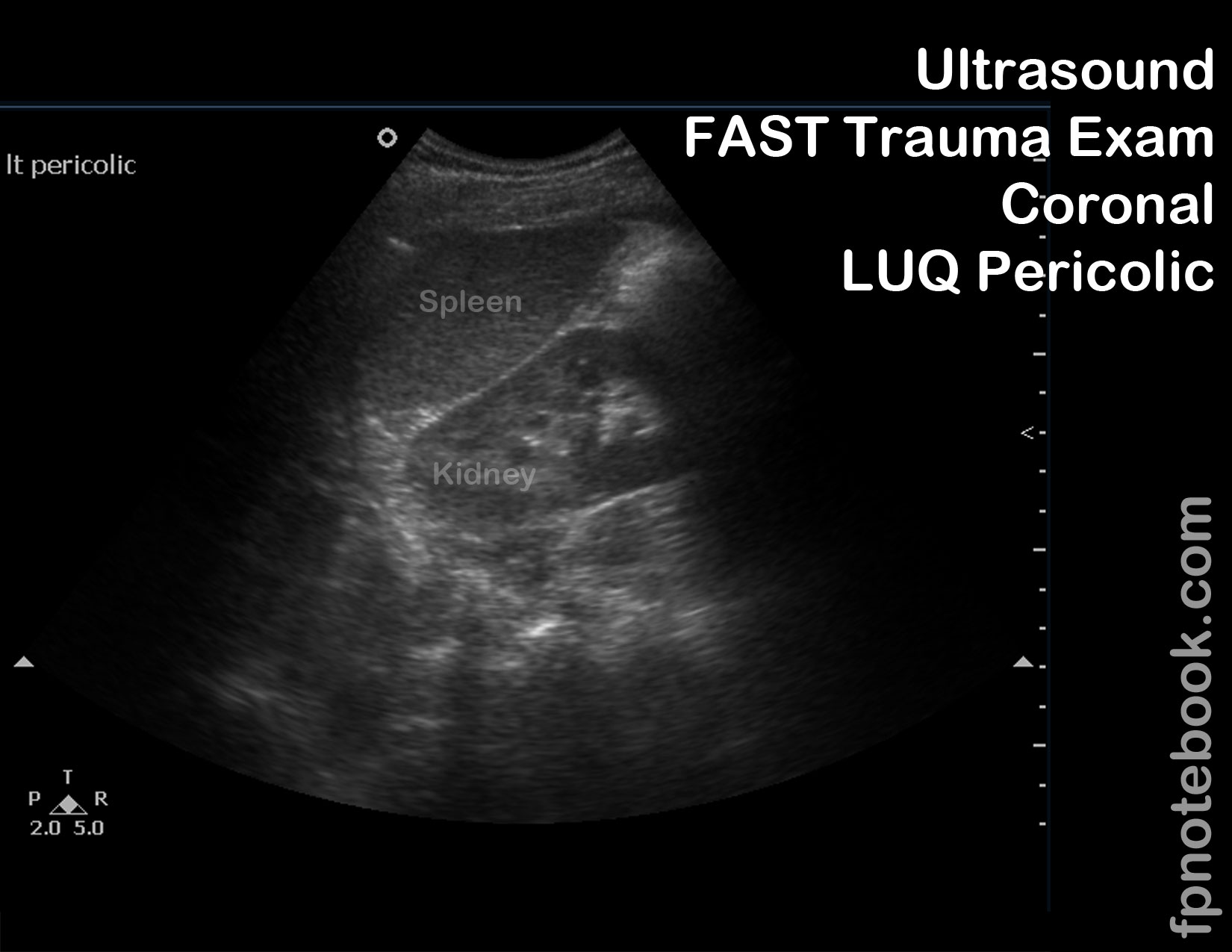
IV. Approach: Left Coronal Ultrasound View
- Obtain view by tilting transducer inferiorly from right intercostal view (or dropping down 1-2 rib spaces)
- Transducer positioning
- Landmarks
- Conditions
- Images

V. Resources
- FAST Exam LUQ (Dr. Mandavia, Sonosite)
VI. References
- Reardon (2016) FAST Scan, Online Video Stabroom.com, accessed 4/1/2016
- Reardon (2013) Emergency Ultrasound Course, 3rd Rock Ultrasound, Minneapolis, MN
- Alameda County Trauma Service FAST Exam
- Mateer (2012) Introduction to Trauma Ultrasound Video, GulfCoast Ultrasound, VL-95-T
- HCMC FAST Exam