
II. Indications
- Anesthesia of the heel and sole of the foot
III. Precautions
- Avoid injecting directly into nerves
- Avoid injection into posterior tibial artery
- Warning: Distal Paresthesias with needle with injection
- Indicates needle is in nerve
- Do not inject here!
- Remove needle and reposition
- Assess degree of Anesthesia
- Wait 10 minutes to assess Anesthetic effect
- If inadequate effect after 10 minutes, may make a second block attempt, or choose alternative Anesthesia
IV. Complications
- Permanent Neuropathy due to posterior tibial nerve injection (do not inject nerve)
V. Preparation
- Needle: 27 gauge 1.5 inch
- Skin Preparation (e.g. Hibiclens or Betadine)
-
Anesthetic
- See Regional Anesthesia for Anesthetic options
- Local Anesthetic 2-5 ml (Ultrasound) or 5-10 ml (landmark)
- Bedside Ultrasound (high frequency linear probe) guidance is recommended
VI. Technique: Sural Nerve Block
- Indications
- Anesthesia for lateral heel and foot
- Landmark Based
- Patient position
- Prone supine, knee flexed and foot flat against the table
- Landmarks
- Lateral border of achilles tendon and
- Proximal to lateral malleolus tip by 1-2 cm
- Inject
- Direct needle posterior to anterior toward the posterior fibula
- After striking fibula, withdraw needle a few mm and aspirate to confirm not intravascular
- Distribute Anesthetic, redirecting needle several times
- Patient position
-
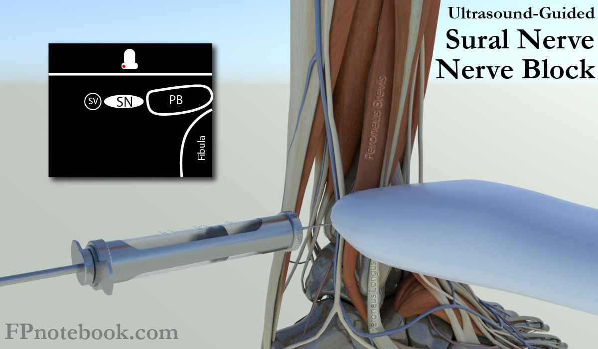
Ultrasound Guided
- See Ankle Ultrasound
- Images
- Patient position
- Lateral decubitus position with lateral ankle up
- Ultrasound Probe
- Linear probe in short axis (transverse) over lateral ankle, proximal to lateral malleolus by 1-3 cm
- Landmarks
- Achilles Tendon (posterior)
- Small Saphenous Vein
- Sural Nerve (adjacent to vein)
- Peroneal Muscles
- Fibula
- Injection
- Insert needle lateral to achilles tendon, directed anteriorly towards lateral malleolus (fibula)
- Inject within the perineural space
VII. Technique: Posterior Tibial Nerve
- Indications
- Anesthesia of majority of heel and sole of foot
- Patient position
- Lateral decubitus position with medial ankle up
- Landmarks
- Tibia (medial malleolus)
- Tibial Artery
- Tibial Nerve
-
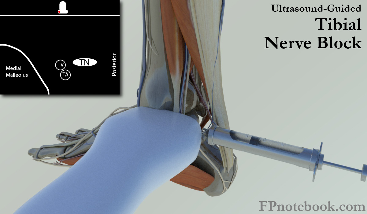
Ultrasound Guided
- Images
- Ultrasound probe
- Linear probe positioned transverse over the medial malleolus (same position as for Saphenous Nerve Block)
- Injection
- Insert needle inline with probe from posterior to anterior
- Tibial nerve is adjacent and posterior to the tibial artery
- Images
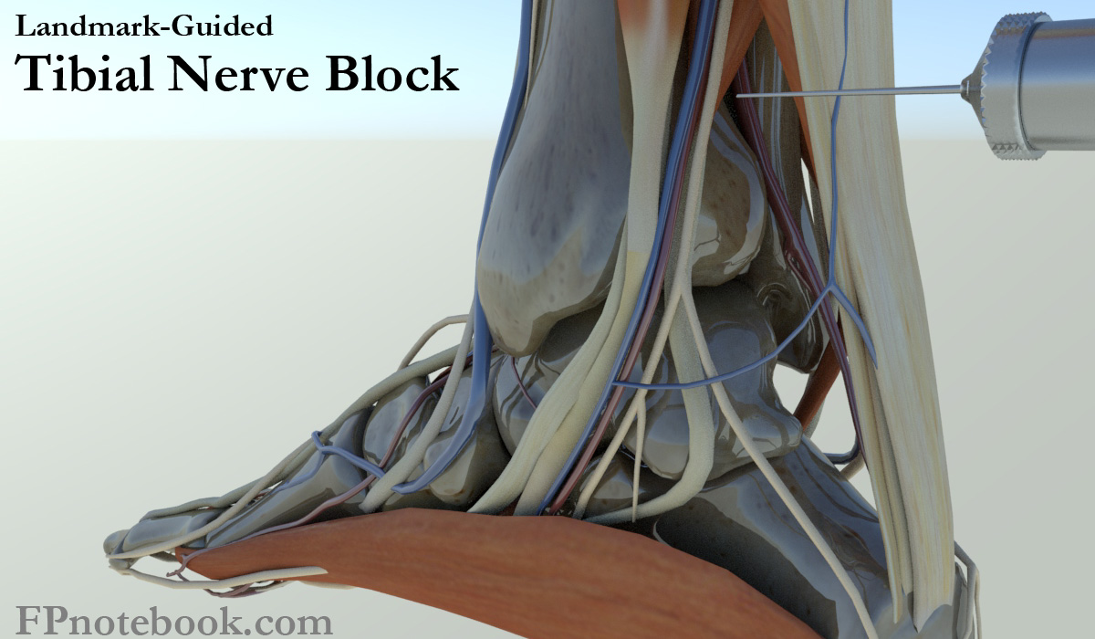
- Landmark Based
- Images
- Insert needle from behind the tibia, 1-2 cm superior (proximal) to the medial malleolus
- Insertion site is anteromedial to the achilles tendon
- Direct needle at the posteromedial tibia (medial malleolus) at a a 45 degree angle
- Needle is inserted and when it strikes the tibia, withdraw the needle back a few mm
- Attempt aspiration confirming not in tibial artery (lies adjacent and anterior to tibial nerve)
- Inject 3 cc
- May inject additional 3-5 cc superficial to nerve while withdrawing needle
- Images
VIII. Resources
IX. References
- Eicken and Rempell (2016) Crit Dec Emerg Med 30(4):3-11
- Pfenninger (1994) Procedures, Mosby, p. 1036-54
- Warrington (2016) Crit Dec Emerg Med 30(7): 12-13
- Salam (2004) Am Fam Physician 69(4):896 [PubMed]
- Yurgil (2020) Am Fam Physician 101(11):654-64 [PubMed]