
II. Images

III. Technique: Anterior (dorsal) Ankle
- Positioning
- Patient lying supine with knee flexed to 90 degrees, foot flat on exam table
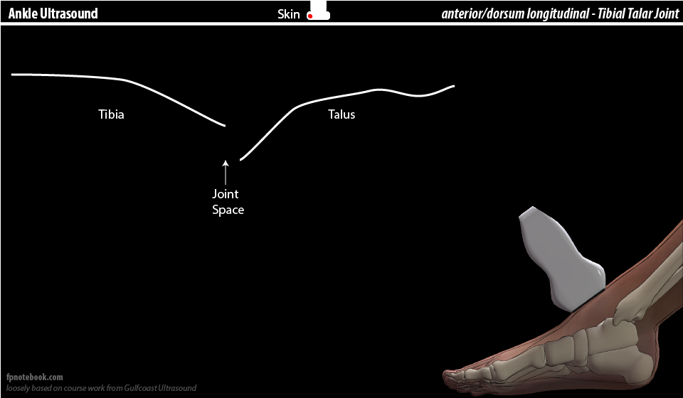
- View 1: Tibia-Talar Joint in Long Axis (LAX)
- Ultrasound probe
- Probe in long axis overlying the tibial talar joint, with the probe indicator toward knee and proximal leg
- Images
- Components (screen left to right)
- Tibia
- Joint space
- Talus (talar dome)
- Ultrasound probe
- View 2: Tibia-Talar Joint in Short Axis (SAX)
- Ultrasound probe
- Rotate probe from LAX (see above) to short axis overlying the joint
- Slide or tilt the probe distally (slight movements) from over the tibia onto the talar dome
- Ultrasound probe
IV. Technique: Lateral Ankle
- Positioning
- Patient lying supine with knee flexed to 90 degrees, foot flat on exam table (same as for anterior ankle)
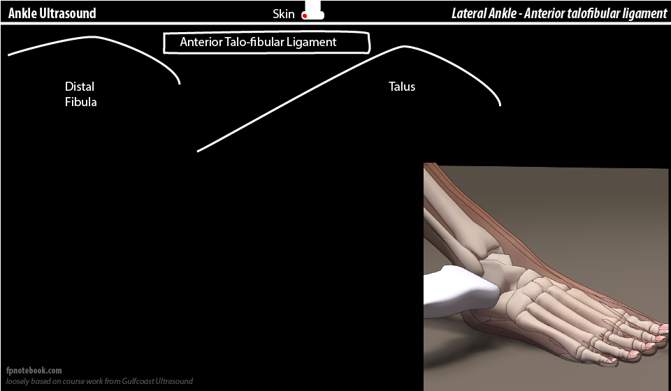
- View 1: Anterior talo-fibular ligament
- Ultrasound probe
- Probe in short axis overlying distal fibula, with the probe indicator toward posterior ankle
- Slide the probe distally from fibula toward talus
- Images
- Components (screen left to right)
- Distal fibula
- Anterior talo-fibular (ATF) ligament
- Talus
- Dynamic maneuvers
- Invert ankle to evaluate integrity of ATF ligament
- Ultrasound probe
- View 2: Peroneal Tendons in long axis (LAX) from posterior-lateral approach
- Ultrasound probe
- Probe in long axis, positioned behind the lateral malleolus and directed posterior to anterior
- Probe indicator toward knee and proximal leg
- Probe may be rotated 90 degrees (to short axis or SAX) to visualize peroneus tendons in cross section
- Components
- Peroneus longus (superficial)
- Attaches ultimately at first Metatarsal base and medial Cuneiform (lateral aspect)
- Peroneus brevis (deeper, immediately deep to longus)
- May be followed around lateral malleolus (rotating 90 degrees) to its attachment at fifth Metatarsal head
- Peroneus longus (superficial)
- Ultrasound probe
V. Technique: Medial Ankle
- Positioning
- Patient lying supine with knee extended and leg externally rotated
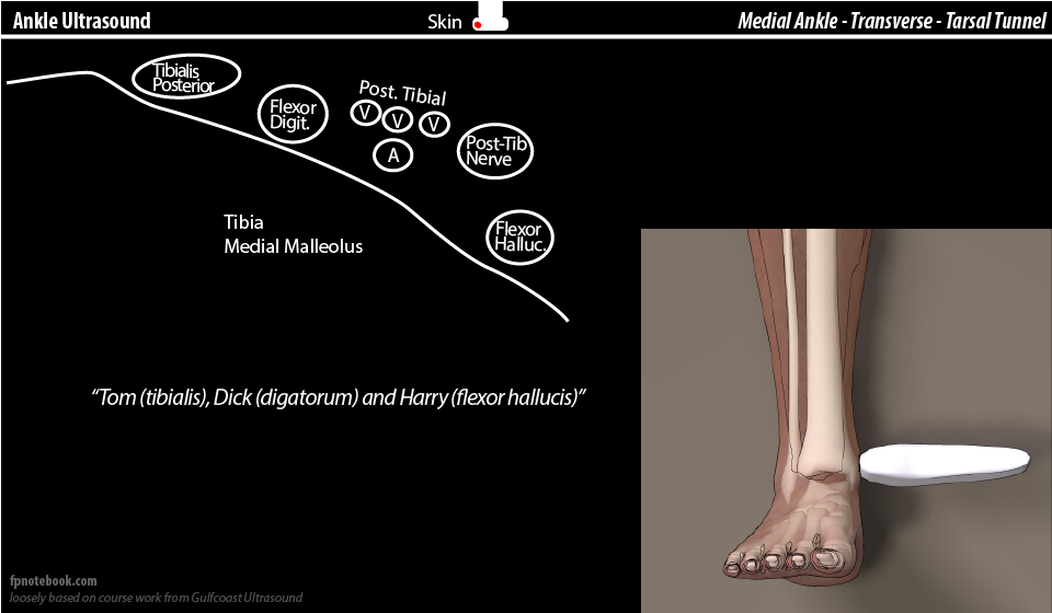
- View 1: Tarsal Tunnel in short axis (SAX)
- Ultrasound probe
- Probe in short axis overlying distal tibia, with the probe indicator toward anterior ankle
- Anterior ankle at screen left and posterior ankle at screen right
- Images
- Components (Mnemonic: "Tom, Dick and Harry")
- Tibialis Posterior tendon (anterior ankle)
- Flexor Digatorum tendon
- Neurovascular bundle
- Posterior tibial veins (several) and one posterior tibial artery
- Posterior tibial nerve
- Flexor hallucis tendon (posterior ankle)
- Tibia (medial malleolus) is deep to these structures
- Ultrasound probe
- View 2: Tarsal Tunnel in long axis (LAX)
- Ultrasound probe
- Rotate probe 90 degrees from SAX view (above)
- Probe in long axis immediately posterior to medial malleolus (distal tibia)
- Probe indicator toward proximal leg (e.g. knee)
- Evaluation areas
- Slide probe posteriorly to identify these structures
- Tibialis Posterior tendon
- Most common cause of Tarsal Tunnel (fluid, swelling may be seen on Ultrasound)
- Posterior tibial nerve
- Lies immediately superficial to the posterior tibial artery
- Posterior tibial artery
- Ultrasound probe
VI. Technique: Posterior Ankle
- Positioning
- Patient lies prone on their Stomache, foot hangs over the end of the bed
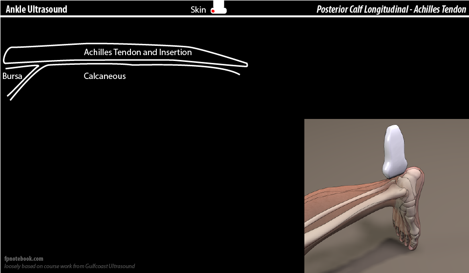
- View 1: Achilles tendon in long axis (LAX)
- Ultrasound probe
- Probe in long axis (LAX) overlying the achilles tendon
- Probe indicator toward proximal leg (e.g. knee)
- Slide Ultrasound probe proximally over the achilles tendon
- Images
- Components: Insertion at Calcaneus
- Achilles Tendon
- Retrocalcaneal Bursa
- Calcaneous (with insertion of achilles tendon)
- Components: Proximal
- Ultrasound probe
- View 2: Achilles tendon in short axis (SAX)
- Ultrasound probe
- Probe in short axis (SAX) overlying the achilles tendon (probe 90 degrees to view above)
- Slide Ultrasound probe distally over the achilles tendon to its calcaneal insertion
- Components
- Ultrasound probe
- View 3: Plantar Fascia in long axis (LAX)
- Ultrasound probe
- Probe in long axis (LAX) overlying the Calcaneus on the plantar foot (slightly medial of center)
- Probe indicator facing up toward posterior foot and ankle
- Components
- Ultrasound probe
VII. References
- Moore (2016) GCUS Musculoskeletal Ultrasound Course, St. Pete's Beach, FL
- Moore (2013) Lower Extremity Ultrasound Video, Gulf Coast Ultrasound