
II. Indications
- Regional Anesthesia of the upper arm, elbow, Forearm and hand
- Incomplete shoulder Anesthesia (must be combined with a higher block for complete shoulder Anesthesia)
III. Complications
- See Regional Anesthesia
- LAST Reaction
- Pneumothorax
- Phrenic Nerve paralysis (transient)
- Common! Do not perform this block bilaterally (risk of respiratory arrest)
- Laryngeal Nerve paralysis (transient)
- Results in transient Hoarseness
- Stellate Ganglion block
IV. Background
- A common perioperative block, known as the "spinal Anesthetic of the arm"
- Originally described in 1911 as a landmark based procedure, and as Ultrasound guided in 1978
V. Landmarks: Ultrasound
- Images
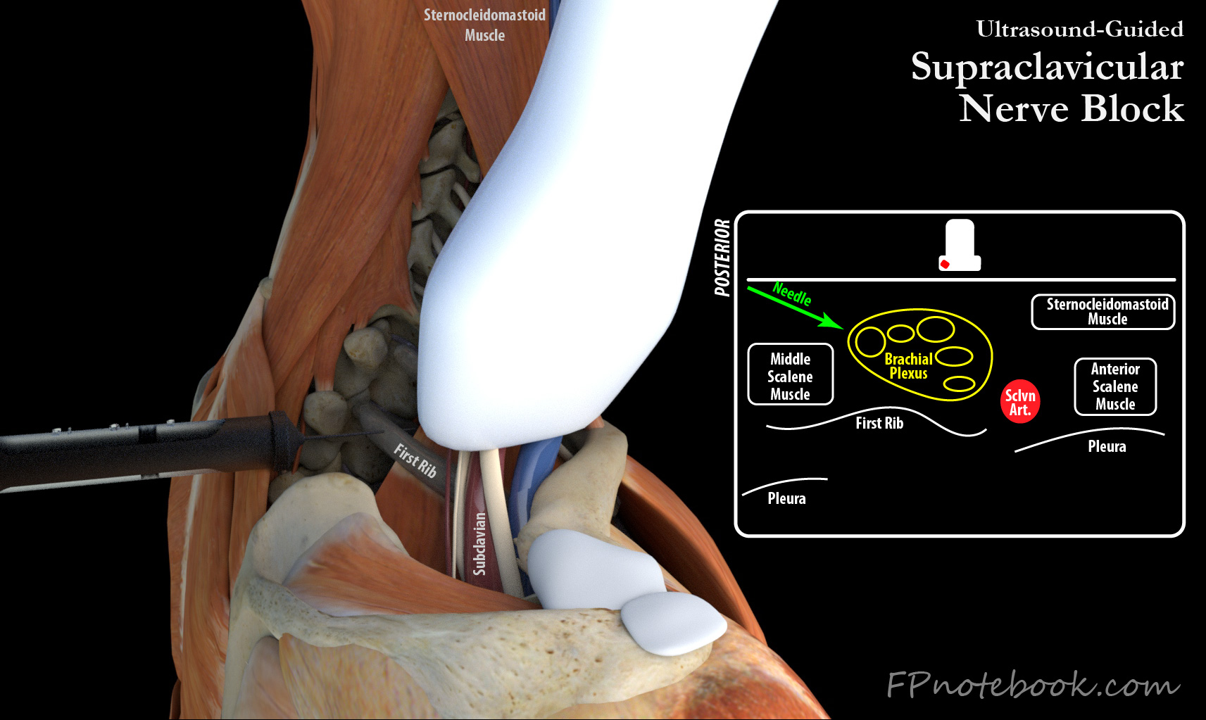
- Linear Ultrasound probe over the lateral neck
- Probe oriented in Transverse Plane, supraclavicular fossa, parallel to the clavicle, directed inferiorly
- Probe slid lateral to internal jugular artery vein
- Indicator at medial neck
- Superficial medial to lateral structures
- Subclavian artery
- Brachial Plexus Trunks ("stop sign")
- Upper Trunk (C5, C6) - superficial
- Middle Trunk (C7)
- Lower or Inferior Trunk (C8, T1) - deepest of nerve trunks
- Neck Muscles
- Deeper medial to lateral structures
- Bones (Clavicle and First Rib)
- First rib is an important landmark to align Ultrasound probe
- Pleura
- Bones (Clavicle and First Rib)
VI. Technique: Ultrasound Guided
- Equipment
- Linear transducer
- Needle 5 to 10 cm, 22 gauge blunt tipped or short bevel Nerve Block needle
- Anesthetic (e.g. Ropivacaine) diluted to 20 to 25 ml
- Patient
- Patient supine at 30 degrees or lateral decubitus with head turned away from the side of the block
- Prepare the skin (Chlorhexidine or Povidone Iodine)
- Needle Insertion
- Ultrasound landmarks as above
- Identify the subclavian artery
- Identify the Brachial Plexus nerve trunks ("stop sign")
- Identify clavicle, and importantly, the first rib which should be in image as a back stop above pleura
- Needle is directed from a lateral approach
- Each level of Brachial Plexus trunk is enveloped in a separate sheath
- Direct needle into lower sheath to anesthetize lower trunk toward first rib (avoid pleura) and inject 10 ml
- Withdraw and Redirect needle between upper and middle sheath and inject 10 ml
- Ultrasound landmarks as above
VII. Monitoring
- Ultrasound guidance with visualization of the needle tip in relation to nerves and vessel is imperative
- Anesthesia also uses nerve stimulators and pressure monitors in this region
VIII. Resources
- Ultrasound-Guided Supraclavicular Brachial PlexusNerve Block (Nysora)
- D'Souza and Johnson (2022) Supraclavicular Block, StatPearls, Trasure Island