
II. Indications
- Anesthesia of Ulnar Nerve distribution distal to wrist (medial hand)
- Consider in combination with Median Nerve Block at Wrist and Radial Nerve Block at Wrist for Complete Hand Anesthesia
III. Contraindications
IV. Anatomy: Landmarks at medial or ulnar wrist
- Ulnar styloid (proximal) or Pisiform Bone (distal)
- Flexor Carpi Ulnaris tendon (medial or ulnar aspect)
- Palpate at region proximal to Pisiform Bone
- Better defined during wrist flexion
- Ulnar Vein
- Ulnar Nerve
- Ulnar Artery (lateral or radial aspect)
V. Precautions
- Avoid injecting directly into Ulnar Nerve
- Avoid injection into ulnar artery
- Confirm adequate Anesthesia at palmar 4th and 5th fingers
VI. Preparation
- Needle: 27 gauge 1.5 inch
- Skin Preparation (e.g. Hibiclens or Betadine)
-
Anesthetic
- See Regional Anesthesia for Anesthetic options
- Local Anesthetic 2-5 ml (Ultrasound) or 5-10 ml (landmark)
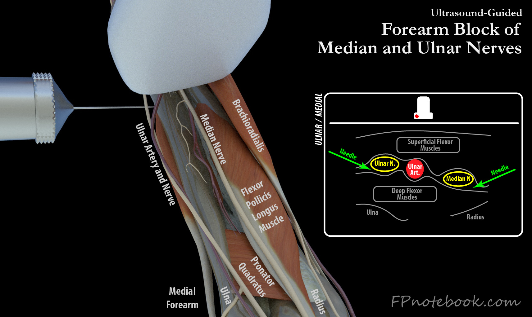
VII. Technique: Forearm (Ultrasound, preferred location compared with wrist)
- Images
-
Ultrasound-Guidance
- High frequency linear probe in short axis
- Use standard Ultrasound-guided Regional Anesthesia technique
- Linear probe is transverse to mid-Forearm
- Median Nerve, Ulnar artery and Ulnar Nerve lie in a plane between the superficial and deep flexors
- Ultrasound at wrist can easily identify ulnar artery and Ulnar Nerve
- Slide probe up Forearm, proximally, until Ulnar Nerve and ulnar artery separate
- Needle Insertion
- Both Ulnar Nerve and Median Nerve can be blocked in same Ultrasound view, with needle redirection
- Approaching from ulnar aspect, rounded Forearm edge allows needle to be perpendicular to probe
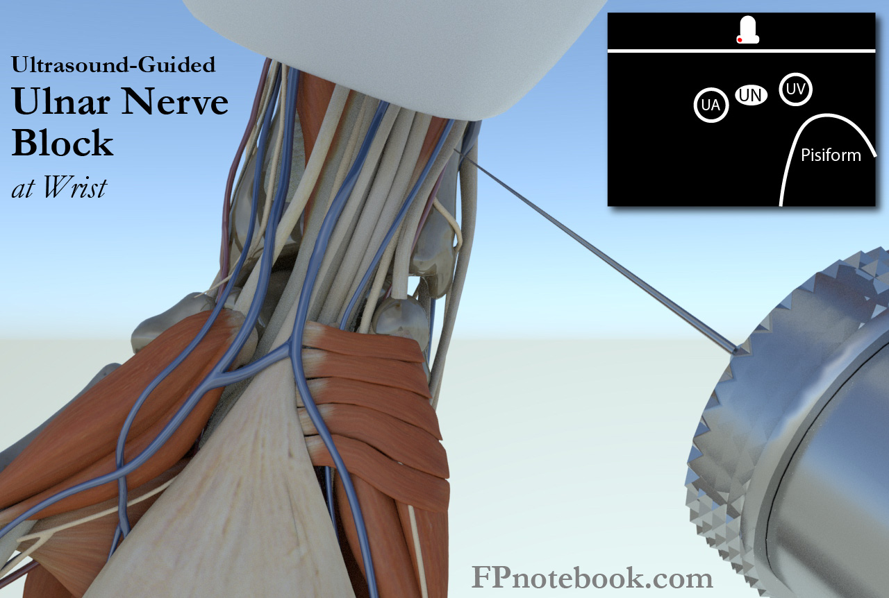
VIII. Technique: Wrist Ultrasound Guided
- Images
- Position
- Patient seated
- Forearm supinated (palm up)
-
Ultrasound Probe Position
- Transverse to Forearm, over the wrist crease
- See Landmarks above
- Injection
- Insert needle at proximal palmar wrist crease
- Direct needle from distal to proximal at 30 degree angle, perpendicular to probe (out-of-plane)
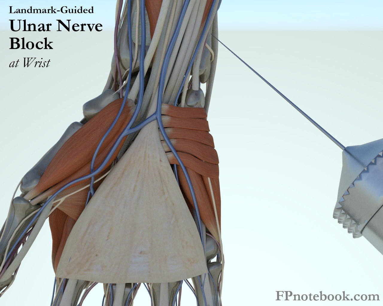
IX. Technique: Landmark
- Images
- Position
- Patient seated
- Forearm supinated (palm up)
- Injection site 1: Palmar branch of the Ulnar Nerve (typical insertion site)
- Direct needle from ulnar aspect of wrist towards radial wrist at 45 degree angle
- Insert at proximal wrist crease (or 1-2 cm proximal), between flexor carpi ulnaris tendon and ulnar artery
- Inject under flexor carpi ulnaris tendon
- Advance needle deeper towards ulna and inject additional 3 ml of Anesthetic while withdrawing needle
- Injection site 2: Dorsal branch of the Ulnar Nerve
- Distal aspect of ulnar styloid process (dorsum)
- Infiltrate 3-4 ml subcutaneously
- Warning: Distal Paresthesias with needle with injection
- Indicates needle is at Ulnar Nerve
- Do not inject here!
- Remove needle and reposition
X. References
- Pfenninger (1994) Procedures, Mosby, p. 1036-54
- Warrington and Sanders (2018) Crit Dec Emerg Med 32(7): 41
- Salam (2004) Am Fam Physician 69(4):896 [PubMed]
- Yurgil (2020) Am Fam Physician 101(11):654-64 [PubMed]