II. Imaging: Views
- Standard
- Anterior-Posterior XRay (or Posterior-Anterior XRay)
- True AP View needed to isolate the two rows of Carpal Bones (oblique view obscures Fractures)
- Evaluate for Scaphoid Fracture, Scapholunate Dissociation and other Carpal BoneFractures
- Lateral XRay
- Anterior-Posterior XRay (or Posterior-Anterior XRay)
- Special
- Scaphoid Fracture suspected
- Scaphoid View
- Pisiform Fracture or hook of hamate Fracture suspected
- Carpal Tunnel view
- Supinated oblique view
- Scapholunate Dissociation
- Clenched-fist view
- Supinated wrist with ulnar deviation
- Scaphoid Fracture suspected
III. Imaging: Other imaging modalities
-
Wrist Ultrasound
- Evaluates soft tissue (tendon, synovium)
- Variable efficacy based on operator
- Bone scan
- Finds occult Fractures (Scaphoid), Stress Fractures
- Highly sensitive but not specific for Fracture
-
Wrist CT Scan
- Identifies Fractures and articular subluxations
-
Wrist MRI
- Identifies Fractures and soft tissue injuries
- Expensive, but most sensitive and specific study
IV. Evaluation: Interpretation
- Post-Reduction Wrist XRay confirms normal radius length
- Images
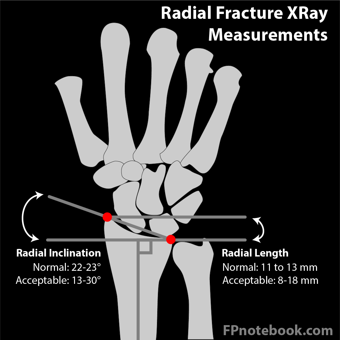
- AP View
- Landmarks on AP View at distal radius
- Distal line (line 1)
- Draw a horizontal line at the level of the radial styloid at the distal radius (point A)
- Represents the distal most point of the radial articular surface
- Proximal line (line 2)
- Draw a horizontal line at the level of the ulnar articulation of the medial distal radius (point B)
- Represents the distal most point of the radial-ulnar articular surface
- Radial length (Radial Height) represents the distance between distal line 1 and proximal line 2
- Articular plane (line 3)
- Draw a line between the points A and B above (between ulnar aspect of radius and ulnar styloid)
- Radial Inclination represents the angle between Line 1 (proximal transverse) and this oblique Line 3
- Distal line (line 1)
- Normal anatomic relationships
- Radial Inclination (normal measurements are for adults)
- Angle formed between the articular plane and the proximal line (see above)
- Normal Radial Inclination: 23.6 +/- 2.5 degrees
- Acceptable inclination: 13-30 degrees
- Radial Height (radial length) shortening (normal measurements are for adults)
- Distance between the proximal and distal lines (see above)
- Normal Radial Height: 11-12 mm
- Acceptable Radial Height: 8-18 mm
- Radial Inclination (normal measurements are for adults)
- Landmarks on AP View at distal radius
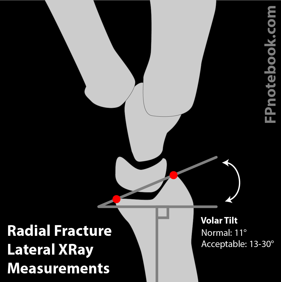
- Lateral View
- Landmarks on Lateral View at distal radius
- Distal dorsal rim
- Point on the distal radius at the dorsal aspect
- Distal volar rim
- Point on the distal radius at the volar aspect
- Vertical line at distal radius
- Perpendicular to the long axis of the radius
- Volar Tilt Line
- Line drawn between distal dorsal rim and distal volar rim
- Distal dorsal rim
- Normal anatomic relationships
- Volar Tilt angle: 11.2 +/- 4.6 degrees
- Angle formed between Volar Tilt line and vertical line at distal radius
- Radius articular surface directed down, forward, in
- Appearance of tea cup and saucer
- Volar Tilt angle: 11.2 +/- 4.6 degrees
- Landmarks on Lateral View at distal radius