
II. Definitions
- Frontal Lobe
- Frontal Lobe is key to cognition, expression, problem solving and memory
- Frontal Lobe lesions may result in Expressive Aphasia, personality changes and Dementia
- The Primary Motor Area (Area 4), as with the Parietal Lobe's sensory regions, is organized into the cortical humunculus
- Disproportionately large region devoted to the face and hands
- Broca's Speech Area (dominant hemisphere, area 44) is key to fluent verbal expression
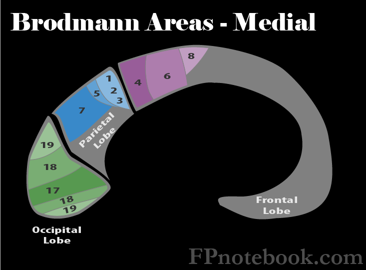
III. Anatomy: Brodmann Areas of Frontal Lobe
- Images
- Primary Motor Area (Area 4)
- Lesions to this area (e.g. CVA) result in Flaccid Paralysis followed by partial recovery
- Babinski Reflex may be present
- Often affected in concert with the lesions to the Supplemental Motor Area (Area 6)
- Supplemental Motor Area (Area 6)
- Lesions often affect Area 4 and Area 6 together, and result in spasticity and hyperreflexia
- Frontal Eye Fields (Area 8)
- Lesions affect voluntary Extraocular Movements looking to the opposite side
- Broca's Speech Area (Area 44, 45)
- Dominant hemisphere lesions to Broca's Speech Area results in motor Aphasia (Broca Aphasia, Expressive Aphasia)
- Patients with motor Aphasia know what they want to say, but their speech is slow and simplified
- Nouns and prepositions are often omitted
- Orbital part of Inferior Frontal Gyrus (Pars Orbitalis, Area 47)
- Involved in the processing of syntax in oral and sign languages, musical syntax, and semantic aspects of language

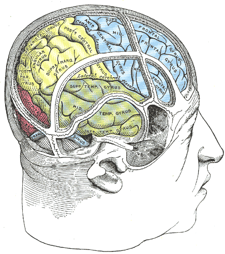
IV. Anatomy: Gyri
- Images
 Lewis (1918) Gray's Anatomy 20th ed (in public domain at Yahoo or BartleBy)
- Precentral Gyrus
- Much of the primary motor cortex corresponds to this gyrus
- Convolution at the posterior Frontal Lobe on the convex side of both Cerebral Hemispheres
- Anterior to the Post Central Gyrus and parallel to the central sulcus
- Central sulcus separates the pre and post central gyri
- Inferior Frontal Gyrus
- Region on the surface of the Frontal Lobe visibly divided in three (pars opercularis, the pars triangularis, Pars Orbitalis)
- Bound by the inferior frontal sulcus dorsally and the lateral fissure ventrally
- Middle Frontal Gyrus
- Large region on the lateral surface of the Frontal Lobe and part of the prefrontal cortex
- Lies between the superior and the inferior frontal sulci and rostral to the Precentral Gyrus
- References
- National Cancer Institute
V. Symptoms: Findings of pathology (e.g. Tumor)
- Personality change
- Expressive Aphasia
VI. Signs: Brain Lesions
- Hemiparesis (Area 4) with or without spasticity and hyperreflexia (Area 6)
- Gait disturbance
- Generalized Seizures or Focal Seizures
- Expressive Aphasia (Areas 44, 45)
- Behavior and Personality change (lesions anterior to the Primary Motor Area, Brodmann 4)
- Judgment and abstract thinking affected
- Affects Instrumental Activities of Daily Living
- May present with concerns for Dementia
VII. Exam: Normal Findings (No lesions)
- Points finger each time examiner makes fist
- Makes fist when examiner points
VIII. References
- Goldberg (2014) Clinical Physiology, Medmaster, Miami, p. 107
- Newton (1994) Am Fam Physician 49(4): 787-97 [PubMed]