
II. Indications
- Carpal Tunnel Syndrome Diagnosis
III. Preparation: Positioning
- Patient sits with Forearm supinated at rest
- Linear, high frequency probe in short axis for the Median Nerve (and the wrist)
- Consider towel roll beneath patient wrist
IV. Images
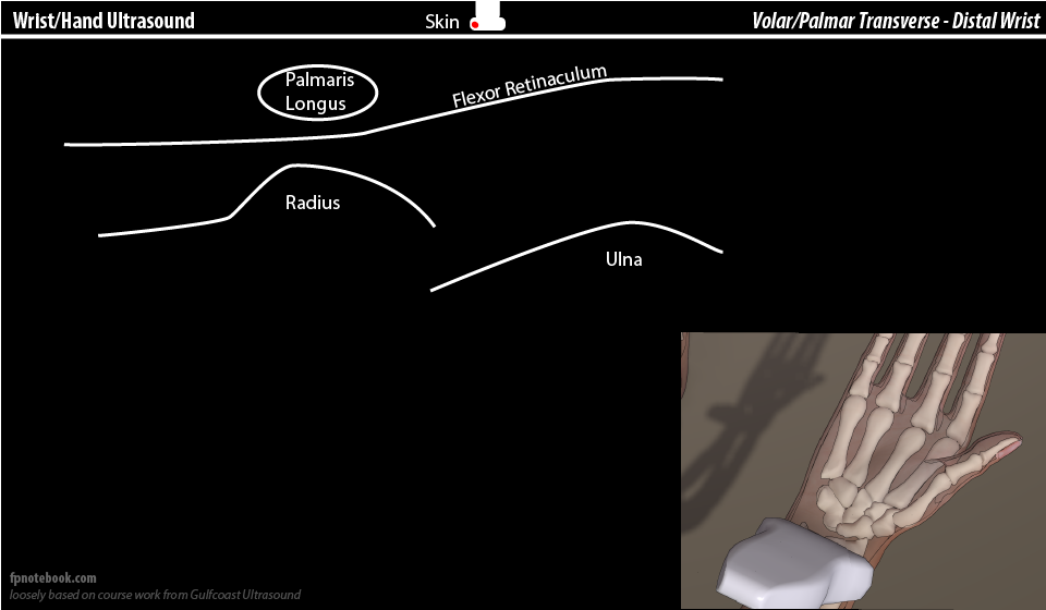
V. Technique: View 1 - Median Nerve at distal wrist
- Landmarks - start position at proximal wrist crease
- Median Nerve is superficial, but within the flexor Retinaculum
- Median Nerve is at radial aspect of palmaris longus
- Median Nerve is adjacent (radial) to flexor pollicis longus
- Wrist adduction and abduction will move flexor pollicis longus and will push on Median Nerve
- Landmarks - measurement position at distal wrist
- Slide probe in SAX distally along volar wrist towards palm
- Goal view
- Flexor Retinaculum most superficial
- Scaphoid shadowing posteriorly at radial or lateral aspect
- Mid-line wrist view contains three main components
- Median Nerve (most superficial)
- Flexor tendons (9)
- Lunate (deepest, shadows posteriorly)
- Pisiform shadowing posteriorly at ulnar or medial aspect
- Precautions
- Avoid compressing Median Nerve with excessive probe pressure
- Biphid Median Nerve or a split Median Nerve (present in >8%)
- Measure each nerve separately and add their areas
- Measurement (in mm2)
- Use the machine cross-sectional area measurement (draw oval around nerve inner boundary)
- Record measurement
VI. Technique: View 2 - Median Nerve at mid-Forearm
- Slide probe proximally (in short axis) from proximal wrist crease, approaching mid-Forearm
- Measurement position is 12 cm from the View 1 measurement
- Landmarks
- Flexor digitorum superficialis
- Median Nerve (within facial layer)
- Improved visualization when patient makes a loose fist
- Flexor digitorum profundus
- Measurement (in mm2)
- As above (draw oval around nerve inner boundary)
VII. Interpretation
-
Wrist to Forearm ratio = (distal wrist median mm2) / (proximal wrist median mm2)
- Ratio >1.4 (some use ratio > 2.0)
- Increase in distal mm2 measurement >2 mm over proximal mm2 measurement
- Proximal Median Nerve width
- Normal if <9 mm2
- Abnormal >12 mm2 (severe if >14 mm2)
- Wrist Median Nerve cross sectional area at Carpal Tunnel inlet
- Cross sectional area >9 mm2 is sensitive and specific for Carpal Tunnel
- Tai (2012) Ultrasound Med Biol 38(7): 1121-8 [PubMed]
VIII. References
- Moore (2016) GCUS Musculoskeletal Ultrasound Course, St. Pete's Beach, FL
- Moore (2013) GCUS Upper Extremity Ultrasound