
II. Epidemiology
- Phalanx Fractures account for 10% of all Fractures and 1-2% of all emergency department visits
- Distal Phalanx Fractures are most common
- Most commonly caused by work injury or Sports Injury
III. Types: Phalanx Fractures
IV. Types: Finger Avulsion Fractures (Tendon Ruptures)
- DIP Extensor Tendon Avulsion (Mallet Finger, Drop Finger, Baseball Finger)
- DIP Flexor Tendon Avulsion (Jersey Finger, Flexor Digitorum Profundus Avulsion)
- PIP Extensor Tendon Avulsion (Central Slip Extensor Tendon Injury, Boutonniere Deformity)
- PIP Flexor Tendon Avulsion (Volar Plate Injury, Jammed Finger, Swan-Neck deformity)
V. Types: Dislocations
VI. Exam
- See Hand Exam (includes Hand Neurovascular Exam)
- Injury exam mantra: "joint above, joint below, circulation, motor function and Sensation, skin and compartments"
- Evaluate for flexor and extensor tendon integrity at MCP, PIP and DIP joints
- Evaluate for rotational alignment (see below)
- Evaluate for open Fracture
VII. XRay
- Hand or finger xray
- Anteroposterior, lateral and oblique views
- Rotational abnormalities may appear on lateral xray as variation in phalanx shaft widths
VIII. Management: General Principles of Hand Fracture Management
- See Epiphyseal Fracture (for Fractures in Children)
- See Interphalangeal Dislocation
- See specific Finger Avulsion Fractures (listed above)
- Nerve Block for any manipulation required at time of Splinting
- Correct angular malalignment and rotation
- Fracture reduction for all unstable, oblique, angulated or displaced Fractures
- Obtain post-reduction xrays after reduction and Splinting
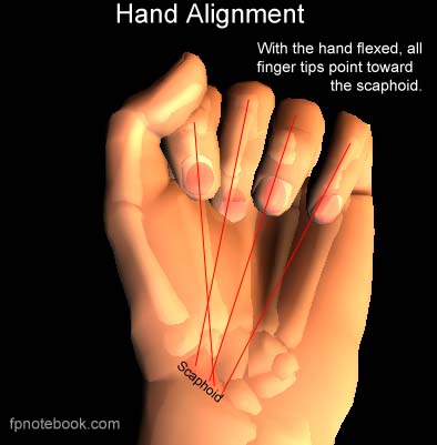
- Axes of all flexed fingers should point toward Scaphoid Bone or radial styloid (thenar eminence)
- Fracture reduction for all unstable, oblique, angulated or displaced Fractures
-
Splinting
- Splint in position of moderate flexion
- Avoid Splinting fingers in extension (esp MCP)
- Avoid over-immobilization (leads to Joint Stiffness and longer recovery)
- Stable, non-displaced Phalanx Fractures may be buddy taped with early protected ROM
- Gutter splint (Ulnar Gutter Splint or Radial Gutter Splint)
- Splint in intrinsic position (30 degrees wrist extension, 90 degrees MCP flexion, IPs in extension)
- Splint in position of moderate flexion
- Evaluate peri-articular Fractures for avulsed tendon
- Avulsed fragments are often attached to a tendon or ligament
- Outpatient follow-up within 5-7 days
- Reevaluate for angulation, rotation or translation
IX. Management: Open Reduction and Internal Fixation (ORIF)
X. References
- Perkins (2020) Crit Dec Emerg Med 34(10): 10-1
- Childress (2022) Am Fam Physician 105(6): 631-9 [PubMed]